4
$\begingroup$

According to Molecular And Cellular Biology (Stephen L. Wolfe),

Membranes disperse almost instantaneously if exposed to a nonpolar environment or to detergents, which are amphipathic molecules that can form a hydrophilic coat around the hydrophobic portions of membrane lipids and proteins in water solutions.

This might be a stupid question but... if detergents can 'form coats around hydrophobic portions' of membrane-suspended molecules, they must, somehow get in the hydrophobic membrane interior... right?

How do they get in the membrane interior? Do they form clusters like endocytic vesicles? What happens after they form hydrophilic coats around hydrophobic molecule regions?

$\endgroup$

2 Answers 2

4
$\begingroup$

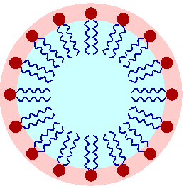
It depends on the concentration, but at higher concentration the detergent molecules build so called micelles, where the hydrophobic "tail" is orientation into the inner part and the hydrophilic "head" is orientated to the outside. This allows the micelle also to fuse with the membrane and then to desintegrate it. This illustration from the Wikipedia shows the schematically:

Detergent micelle in water

Its from the surfactant article, which has more details.

$\endgroup$
3
$\begingroup$

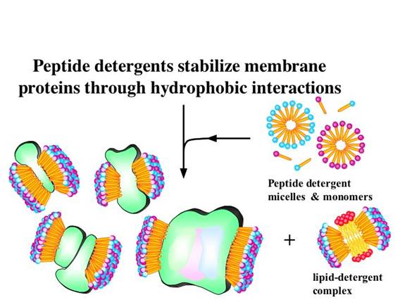
The membrane is dispersed by detergents. But the detergents, under the right concentrations and conditions (salt, pH, et) form micelles with smaller curvature than the lipids that compose cell membranes. With some luck, they can form a small hydrophobic micelle bubble around the protein.

This figure is depicts detergent micelles around a membrane protein's membrane bound regions, hopefully without distorting the shape of the protein!

enter image description here

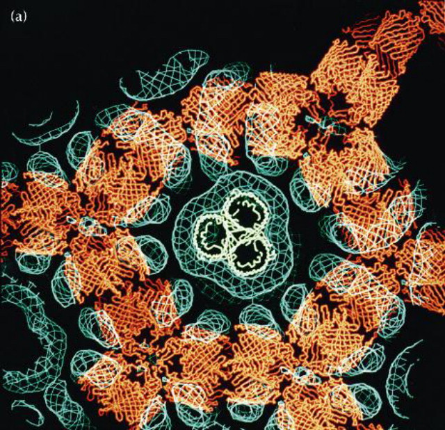
Since you asked in the comments, here is a picture of the electron density of the detergent micelle in ompF Porin crystals - the detergent density is blue the protein is red. For a while in the mid 90s through maybe a few years ago there were a reasonable number of simulations including detergent molecules. The mobility of the chains in the micelles means that no specific model structure would be accurate, but it can help understand the electromagnetic environment of the solvent in cases which are important like charge transfer in a photoreaction center or the dynamics of transport through an import/export protein.

enter image description here

$\endgroup$
4
  • 1
    $\begingroup$ Thats a great image. $\endgroup$
    – Chris
    Commented Dec 24, 2013 at 12:59
  • $\begingroup$ thanks. If you want to see a more true to life one, many membrane proteins have visualized detergent micelles in simulations and in the crystal unit cell. I'll add another image above. $\endgroup$
    – shigeta
    Commented Dec 25, 2013 at 3:29
  • $\begingroup$ sorry... haven't been in SE for months; and DO please show me that image! @shigeta $\endgroup$
    – hello all
    Commented Feb 17, 2014 at 14:43
  • $\begingroup$ actually the last image there is a good example... the blue density is all micelle in a porin unit cell. protein is red $\endgroup$
    – shigeta
    Commented Feb 17, 2014 at 15:37

You must log in to answer this question.

Not the answer you're looking for? Browse other questions tagged .