Excitatory neurons in layer 4 are not all stellate in all cortical areas, for example see Smith and Populin, 2001 who show clearly that in auditory cortex most excitatory cells in layer 4 are pyramidal. There are also many cortical areas that have no clear "layer 4" and also do not contain spiny stellate cells.
In general, intracortical circuitry is not fully understood. Although your question shows understanding of this next point, let me highlight it anyways because it is a point that is often forgotten: neither axons nor dendrites are constrained to the layers containing soma. Therefore, it is possible for a cell of "layer A" to contact a cell of "layer B" even if the "layer A" cell has no axons in "layer B" and the cell of "layer B" has no dendrites in "layer A": they could have axons and dendrites in "layer C", for example.
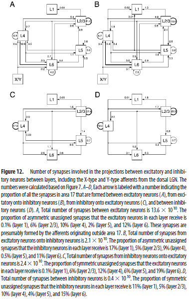
One approach has been taken by some such as Binzegger et al., 2004 is to estimate intralaminar cortical connectivity based on co-occurrence within a given layer of axons and dendrites. This approach is an approximation, of course, because there is no guarantee that axons contact dendrites randomly within a given layer. For example, axons could preferentially contact the dendrites of inhibitory rather than excitatory cells, and this would fundamentally influence the circuit dynamics.
The result from Binzegger et al (their Figure 1 is a fairly clear summary figure, if you want to start there) is that, although there are some predominant patterns of connectivity, there is no pure feed-forward circuit: all of the connections are at least partially reciprocal. For the example you gave, layer 2/3 to layer 4, the number of connections from layer 2/3 to layer 4 is estimated to be about half as many as the number of connections from layer 4 to layer 2/3.

Figure from Binzegger et al, 2004; Creative Commons Attribution-Noncommercial-Share Alike 3.0 Unported License
However, it is also possible that these connections preferentially target inhibitory cells rather than excitatory cells in layer 4: see Thomson et al., 2002.
It is likely that the variety in sources/diagrams you are familiar with is simply because people take different approaches to complex systems (and different approaches may be warranted in different situations). Some people choose to highlight the most salient features, perhaps the strongest layer-to-layer connections, in the hope that this produces an understandable, though simplified, model. Other people choose to highlight the vast complexity, emphasizing all of the smaller details that may actually be important.
For an analogy, a chef is likely to just deal with olive oil. They don't care that olive oil is actually a collection of several fatty acids: it's just olive oil to them, and the importance to them is that olive oil is distinct from another vegetable oil. A biochemist, however, might be interested in the specific fatty acids and might point out that those specific fatty acids are found in all sorts of other oils. Neither one is likely to convince the other that they have the better answer for all purposes.
Neither approach is strictly incorrect or better than the other, until better understanding makes one of the approaches obsolete.
References:
Binzegger, T., Douglas, R. J., & Martin, K. A. (2004). A quantitative map of the circuit of cat primary visual cortex. Journal of Neuroscience, 24(39), 8441-8453.
Smith, P. H., & Populin, L. C. (2001). Fundamental differences between the thalamocortical recipient layers of the cat auditory and visual cortices. Journal of Comparative Neurology, 436(4), 508-519.
Thomson, A. M., West, D. C., Wang, Y., & Bannister, A. P. (2002). Synaptic connections and small circuits involving excitatory and inhibitory neurons in layers 2–5 of adult rat and cat neocortex: triple intracellular recordings and biocytin labelling in vitro. Cerebral cortex, 12(9), 936-953.

