7
$\begingroup$

The transporters in the plasma membrane of the cells promote the entry of glucose molecules from the extracellular matrix to the cytosol of the cell. Could someone explain how does the nutrient molecule enter the extracellular space from the blood vessel?

For instance, in the context of the pancreas, the walls of the blood vessel is fenestrated. The literature also provides evidence for the presence of connexon in the endothelium of the capillaries.

My doubt is, the nutrient molecule that diffuses from the blood vessel reaches the cytosol of the cell through

  1. Diffusing through connexon ?(or)
  2. Does it reach the interstitial matrix(the fluid surrounding the cells) and then uptaken by the transporters present in the plasma membrane of the cell?
$\endgroup$
2
  • $\begingroup$ Connexons form gap junctions right? I am not sure because right now I don't have any appropriate literature to site for good answer but doesn't glucose transport involve solely GLUT? I mean if connexons were the option then why would diabetes even happen?Please do rectify me if I am going wrong. $\endgroup$
    – user 33690
    Commented Sep 24, 2018 at 19:56
  • $\begingroup$ @user33690 Yes, connexons form gap junctions. GLUT transporters are involved in the active transport of glucose molecules from the extracellular space. For example, in the liver, the capillaries(sinusoids) are surrounded by the perisinusoidal space, the cells are found adjacent to the perisinusoidal space. Likewise, is there a layer, surrounding the fenestrated capillaries, from which the nutrients can enter the cell? My question is, which is the region in the pancreas that supplies nutrients to the surface of the cells? Any further thoughts? $\endgroup$
    – Natasha
    Commented Sep 25, 2018 at 3:53

1 Answer 1

4
$\begingroup$

I think I understand your question, Natasha. In short, your own answer #2 is correct.

There are 3 spaces, and 2 pathways for glucose to pass from one to the next:

  1. intracapillary plasma
  2. extracellular fluid
  3. the cytosol.

Ways glucose gets into the cell:

  • (2->3) To get from the ECF to the cytosol , glucose always needs a transport protein. These are the GLUTs. In two cases, the small intestine and kidney, these are part of a secondary active transport system based on the Na/K-ATPase. In the pancreas, it's GLUT2.

  • (1->2) To get from the capillary plasma to the ECF requires filtration, the process of applying hydrostatic pressure to the plasma and literally squeezing it like a sponge. The boundary of the "blood sponge" is the basement membrane. The membrane holds in the proteins, and lets anything dissolved in the watery serum (like glucose) through.

The Filtration Constant Kf is proportional to the percentage of the BM that is exposed in a given capillary, which varies by the type and other factors like histamine release. enter image description here Type 1 (continuous) has the lowest exposure of the BM (only the intercellular clefts). Type 2 (fenestrated)has the clefts and the fenestrations to expose the BM. Type 3 (sinusoids) have huge gaps between the cells, and importantly, an incompetent BM that allows proteins and cells through along with the watery serum.
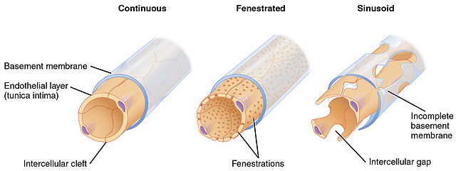
Dynamic factors that change filtration rates:

  • Histamine causes the post-capillary venule's endothelial cells to contract, and exposes more of the BM allowing more serum filtration, but it also allows neutrophil extravasation, during which the neutrophils punch holes in the BM through which white cells can squeeze, which is why you get a proteinaceous exudate in Type I hypersensitivity.

  • Increase in blood flow Like a water balloon increasing its surface area with more blood flow, skeletal muscle capillary beds (Type I) increase their volume and surface area when engorged with blood during exercise. This doesn't change the Kf (the only exit for serum is still the intercellular clefts, but they're bigger now).

P.S. A 1985 morphometric study of pancreatic capillaries shows that the idea that endocrine glands need a boatload of bloodflow (high serum filtration) is valid. Even within a single capillary that feeds both an exocrine acinus and and endocrine islet, the side facing the endocrine islet had more fenestrae.

$\endgroup$
10
  • 1
    $\begingroup$ From what I understand from the above explanation, the BM covers the endothelial layer .When the plasma is filtered out of the pores in the endothelial layer of the fenestrated capillary, how does it cross the BM? Will there be pores in the BM too? $\endgroup$
    – Natasha
    Commented Sep 27, 2018 at 4:57
  • $\begingroup$ Could you please provide references of studies that measure the Starling forces in the capillaries of the endocrine glands of the pancreas, if you have come across these studies in the past? I could only find the measurements of hydrostatic pressure and osmotic pressure in the intestine of cat.I have been looking for these measurements in the capillaries that pass through islets. $\endgroup$
    – Natasha
    Commented Sep 27, 2018 at 5:09
  • 1
    $\begingroup$ Terms to understand filtration. Blood = cells + plasma. Plasma = proteins + serum. Serum = water + ions + small-molecule solutes. Due to the transmural pressure in the capillary, the blood gets squeezed. Stuff that is too big to make it across the filtration barrier gets left inside (cells + proteins). The plasma gets filtered, like spaghetti in a colander. The proteins/spaghetti stay inside, and the watery serum gets through because it's small enough. The BM is made of Type IV and other collagens, laminins, and other adhesion PROTEINS. (more in next comment, ran out of room) $\endgroup$
    – vipatron
    Commented Sep 27, 2018 at 12:06
  • 1
    $\begingroup$ All proteins get their 3-d shape because they are dissolved in water. It is everywhere in the body. It surrounds each protein, and fills the space between them unless adjacent proteins have hydrophobic pockets for protein-protein interactions. So, even though there aren't pores you can see on EM, there are gaps to accomodate bulk flow (called convective flow). To answer your second question, that's a pretty specific study you're asking for, and I haven't read it. Have you tried a PubMed search? $\endgroup$
    – vipatron
    Commented Sep 27, 2018 at 12:09
  • $\begingroup$ +1 "The plasma gets filtered, like spaghetti in a colander".Couldn't have explained it better!! If my understanding is correct, in a fenestrated capillary, there is convection in the inner channel, filtration across the inner channel, and convection in the space between the inner channel and the BM.A silly doubt, is the fluid between the inner channel and BM referred to as the interstitial fluid? How do the cells get in contact with this fluid? $\endgroup$
    – Natasha
    Commented Sep 28, 2018 at 4:52

You must log in to answer this question.

Not the answer you're looking for? Browse other questions tagged .