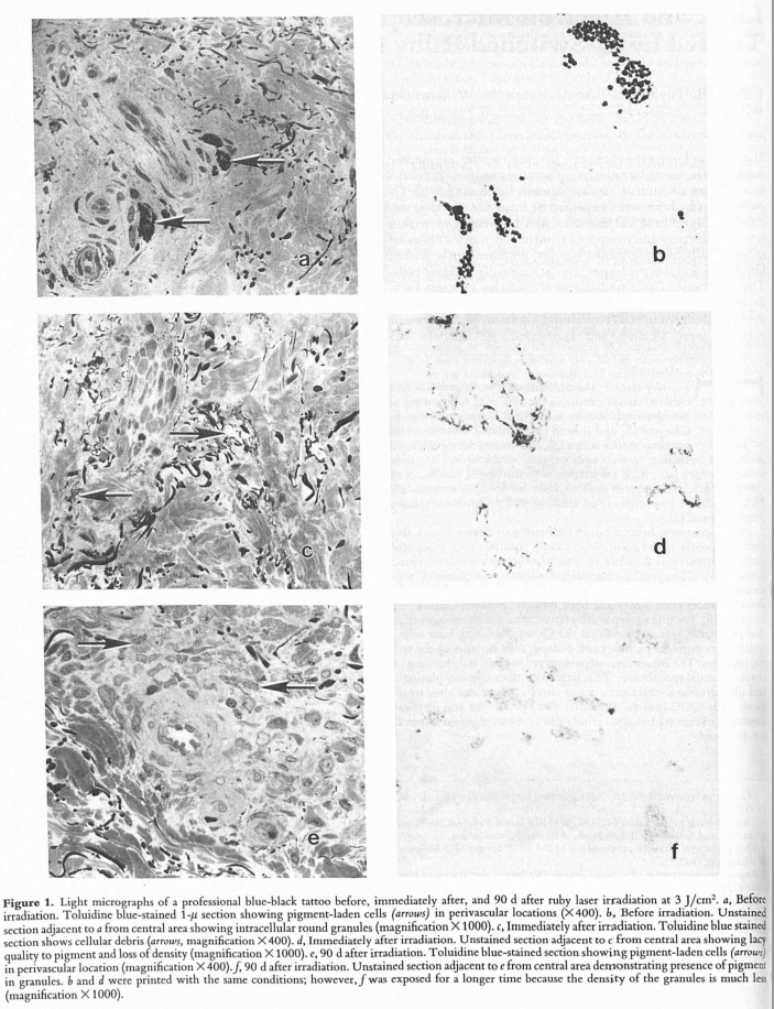
Contrary to the other answer posted, this paper shows via microscope images that the tattoo ink is in fact absorbed into cells, and forms small intracellular round granules.
Electron microscopy of untreated tattoos revealed membrane-bound pigment granules, predominantly within fibroblasts and macrophages, and occasionally in mast cells. These granules contained pigment particles.
 Images a, c and e are images of toluidine blue stained cells, while images b, d and f are images of unstained cells showing the tattoo pigments.
Images a and b are prior to the laser treatment, c and d directly after the treatment, and e and f 90 days post treatment with the laser.
The reason why the tattoo marks persist is not because the pigments are deposited extracellularly, but that they are deposited intracellularly.
The pigments form intracellular granules that are not broken down, and therefore in the absence of external forces such as a laser, the pigments will remain there for long periods of time.
This paper (thanks to biozic) also describes the tattoo ink being persistently found intracellularly instead of extracellularly.
Biopsies obtained from tattoos 1, 2, 3, and 40 years old differed only in the types of ink used. All the ink particles were found to be located in dermal cells. The epidermis was completely devoid of pigment particles. The basement membrane was continuous at the epidermal-dermal junction. Ink particles were found throughout the upper dermis but all were within the boundary of a cell membrane.
According to this TED video (which unfortunately does not state its primary sources), the fibroblasts that contain these engulfed ink particles are themselves taken up by newer fibroblasts when they die, therefore the ink particles remain in the dermis and are not removed by cellular renewal.
