Added a rider explaining why diagrams of beta-sheets are the way they are.
Source Link
David
  • 26.6k
  • 8
  • 53
  • 95

This sort of question is easily answered by consulting and carefully reading an introductory biochemistry text, like this section of Berg et al. on line. However, as the diagrams of β-sheets can be confusing, I will summarize.

  • The amino acids linked by covalent peptide bonds produce a protein molecule in the form of a single polypeptide chain.
  • This polypeptide chain folds into a specific shape in three-dimensional space, its conformation being maintained by weak non-covalent interactions — ionic bonds, hydrogen bonds and Van der Walls interactions. (Often referred to as protein tertiary structure.) Proteins that have a roughly spheroidal shape (rather than being long and extended) are referred to as globular proteins.
  • Often the three-dimensional structure has large regular components (often referred to as secondary structure) because of the potential for repeated hydrogen bonding between the CO and NH of the repeating peptide bonds.

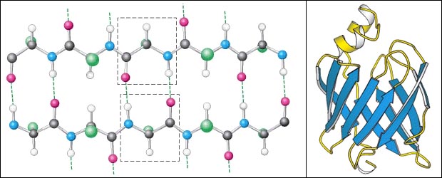
Molecular diagrams to illustrate the type of secondary structure known as the β-sheet generally show just a section of the polypeptide chain for clarity, as in the left-hand illustration from Berg et al., below:

Protein secondary structure

This may give the impression that two separate chains are involved†, but cartoon representations such as the one on the right (also from the same section of Berg et al.) show that for globular proteins this is not so.

In the diagram the flat blue arrows represent β-strands that are hydrogen-bonded to one another to form a twisted barrel-shaped β-sheet. The yellow ‘rope’ between the arrows represents the continuation of the chain in the non-sheet areas of the structure.

So the possibility for confusion arises primarily from the difficulty of showing the detailed pattern of hydrogen bonding in the whole of a large protein. (Although it may also arise from the fact that it is easiest to illustrate β-sheets using the diagrams of silk fibroin — historically the first to be described — in which the sheets are very regular and flat. To an extent this is misleading as the right-hand side of my diagram indicates.)

Footnote

Fibrous proteins, such as silk, exist in which β-sheets are formed between many different chains. The identification of the individual chains can be made unequivocally, e.g. on the basis of the gene sequence, and it is clear that the higher-order structure is composed of many individual molecules. In smaller higher-order structures, e.g. tetramers of globular proteins held together by weak non-covalent interactions, the semantic question of whether a molecule can be made up of several individual chains arises. In my opinion, this question is best ignored, unless one is studying the biophysical chemistry of proteins.

This sort of question is easily answered by consulting and carefully reading an introductory biochemistry text, like this section of Berg et al. on line. However, as the diagrams of β-sheets can be confusing, I will summarize.

  • The amino acids linked by covalent peptide bonds produce a protein molecule in the form of a single polypeptide chain.
  • This polypeptide chain folds into a specific shape in three-dimensional space, its conformation being maintained by weak non-covalent interactions — ionic bonds, hydrogen bonds and Van der Walls interactions. (Often referred to as protein tertiary structure.) Proteins that have a roughly spheroidal shape (rather than being long and extended) are referred to as globular proteins.
  • Often the three-dimensional structure has large regular components (often referred to as secondary structure) because of the potential for repeated hydrogen bonding between the CO and NH of the repeating peptide bonds.

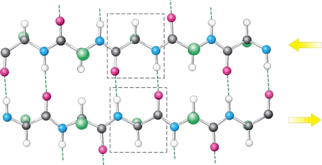
Molecular diagrams to illustrate the type of secondary structure known as the β-sheet generally show just a section of the polypeptide chain for clarity, as in the left-hand illustration from Berg et al., below:

Protein secondary structure

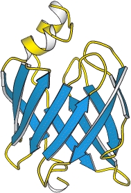
This may give the impression that two separate chains are involved†, but cartoon representations such as the one on the right (also from the same section of Berg et al.) show that for globular proteins this is not so.

In the diagram the flat blue arrows represent β-strands that are hydrogen-bonded to one another to form a twisted barrel-shaped β-sheet. The yellow ‘rope’ between the arrows represents the continuation of the chain in the non-sheet areas of the structure.

So the possibility for confusion arises primarily from the difficulty of showing the detailed pattern of hydrogen bonding in the whole of a large protein.

Footnote

Fibrous proteins, such as silk, exist in which β-sheets are formed between many different chains. The identification of the individual chains can be made unequivocally, e.g. on the basis of the gene sequence, and it is clear that the higher-order structure is composed of many individual molecules. In smaller higher-order structures, e.g. tetramers of globular proteins held together by weak non-covalent interactions, the semantic question of whether a molecule can be made up of several individual chains arises. In my opinion, this question is best ignored, unless one is studying the biophysical chemistry of proteins.
This sort of question is easily answered by consulting and carefully reading an introductory biochemistry text, like this section of Berg et al. on line. However, as the diagrams of β-sheets can be confusing, I will summarize.

  • The amino acids linked by covalent peptide bonds produce a protein molecule in the form of a single polypeptide chain.
  • This polypeptide chain folds into a specific shape in three-dimensional space, its conformation being maintained by weak non-covalent interactions — ionic bonds, hydrogen bonds and Van der Walls interactions. (Often referred to as protein tertiary structure.) Proteins that have a roughly spheroidal shape (rather than being long and extended) are referred to as globular proteins.
  • Often the three-dimensional structure has large regular components (often referred to as secondary structure) because of the potential for repeated hydrogen bonding between the CO and NH of the repeating peptide bonds.

Molecular diagrams to illustrate the type of secondary structure known as the β-sheet generally show just a section of the polypeptide chain for clarity, as in the left-hand illustration from Berg et al., below:

Protein secondary structure

This may give the impression that two separate chains are involved†, but cartoon representations such as the one on the right (also from the same section of Berg et al.) show that for globular proteins this is not so.

In the diagram the flat blue arrows represent β-strands that are hydrogen-bonded to one another to form a twisted barrel-shaped β-sheet. The yellow ‘rope’ between the arrows represents the continuation of the chain in the non-sheet areas of the structure.

So the possibility for confusion arises primarily from the difficulty of showing the detailed pattern of hydrogen bonding in the whole of a large protein. (Although it may also arise from the fact that it is easiest to illustrate β-sheets using the diagrams of silk fibroin — historically the first to be described — in which the sheets are very regular and flat. To an extent this is misleading as the right-hand side of my diagram indicates.)

Footnote

Fibrous proteins, such as silk, exist in which β-sheets are formed between many different chains. The identification of the individual chains can be made unequivocally, e.g. on the basis of the gene sequence, and it is clear that the higher-order structure is composed of many individual molecules. In smaller higher-order structures, e.g. tetramers of globular proteins held together by weak non-covalent interactions, the semantic question of whether a molecule can be made up of several individual chains arises. In my opinion, this question is best ignored, unless one is studying the biophysical chemistry of proteins.
fixed typo
Source Link
David
  • 26.6k
  • 8
  • 53
  • 95

This sort of question is easily answered by consulting and carefully reading an introductroyintroductory biochemistry text, like this section of Berg et al. on line. However, as the diagrams of β-sheets can be confusing, I will summarize.

  • The amino acids linked by covalent peptide bonds produce a protein molecule in the form of a single polypeptide chain.
  • This polypeptide chain folds into a specific shape in three-dimensional space, its conformation being maintained by weak non-covalent interactions — ionic bonds, hydrogen bonds and Van der Walls interactions. (Often referred to as protein tertiary structure.) Proteins that have a roughly spheroidal shape (rather than being long and extended) are referred to as globular proteins.
  • Often the three-dimensional structure has large regular components (often referred to as secondary structure) because of the potential for repeated hydrogen bonding between the CO and NH of the repeating peptide bonds.

Molecular diagrams to illustrate the type of secondary structure known as the β-sheet generally show just a section of the polypeptide chain for clarity, as in the left-hand illustration from Berg et al., below:

Protein secondary structure

This may give the impression that two separate chains are involved†, but cartoon representations such as the one on the right (also from the same section of Berg et al.) show that for globular proteins this is not so.

In the diagram the flat blue arrows represent β-strands that are hydrogen-bonded to one another to form a twisted barrel-shaped β-sheet. The yellow ‘rope’ between the arrows represents the continuation of the chain in the non-sheet areas of the structure.

So the possibility for confusion arises primarily from the difficulty of showing the detailed pattern of hydrogen bonding in the whole of a large protein.

Footnote

Fibrous proteins, such as silk, exist in which β-sheets are formed between many different chains. The identification of the individual chains can be made unequivocally, e.g. on the basis of the gene sequence, and it is clear that the higher-order structure is composed of many individual molecules. In smaller higher-order structures, e.g. tetramers of globular proteins held together by weak non-covalent interactions, the semantic question of whether a molecule can be made up of several individual chains arises. In my opinion, this question is best ignored, unless one is studying the biophysical chemistry of proteins.

This sort of question is easily answered by consulting and carefully reading an introductroy biochemistry text, like this section of Berg et al. on line. However, as the diagrams of β-sheets can be confusing, I will summarize.

  • The amino acids linked by covalent peptide bonds produce a protein molecule in the form of a single polypeptide chain.
  • This polypeptide chain folds into a specific shape in three-dimensional space, its conformation being maintained by weak non-covalent interactions — ionic bonds, hydrogen bonds and Van der Walls interactions. (Often referred to as protein tertiary structure.) Proteins that have a roughly spheroidal shape (rather than being long and extended) are referred to as globular proteins.
  • Often the three-dimensional structure has large regular components (often referred to as secondary structure) because of the potential for repeated hydrogen bonding between the CO and NH of the repeating peptide bonds.

Molecular diagrams to illustrate the type of secondary structure known as the β-sheet generally show just a section of the polypeptide chain for clarity, as in the left-hand illustration from Berg et al., below:

Protein secondary structure

This may give the impression that two separate chains are involved†, but cartoon representations such as the one on the right (also from the same section of Berg et al.) show that for globular proteins this is not so.

In the diagram the flat blue arrows represent β-strands that are hydrogen-bonded to one another to form a twisted barrel-shaped β-sheet. The yellow ‘rope’ between the arrows represents the continuation of the chain in the non-sheet areas of the structure.

So the possibility for confusion arises primarily from the difficulty of showing the detailed pattern of hydrogen bonding in the whole of a large protein.

Footnote

Fibrous proteins, such as silk, exist in which β-sheets are formed between many different chains. The identification of the individual chains can be made unequivocally, e.g. on the basis of the gene sequence, and it is clear that the higher-order structure is composed of many individual molecules. In smaller higher-order structures, e.g. tetramers of globular proteins held together by weak non-covalent interactions, the semantic question of whether a molecule can be made up of several individual chains arises. In my opinion, this question is best ignored, unless one is studying the biophysical chemistry of proteins.

This sort of question is easily answered by consulting and carefully reading an introductory biochemistry text, like this section of Berg et al. on line. However, as the diagrams of β-sheets can be confusing, I will summarize.

  • The amino acids linked by covalent peptide bonds produce a protein molecule in the form of a single polypeptide chain.
  • This polypeptide chain folds into a specific shape in three-dimensional space, its conformation being maintained by weak non-covalent interactions — ionic bonds, hydrogen bonds and Van der Walls interactions. (Often referred to as protein tertiary structure.) Proteins that have a roughly spheroidal shape (rather than being long and extended) are referred to as globular proteins.
  • Often the three-dimensional structure has large regular components (often referred to as secondary structure) because of the potential for repeated hydrogen bonding between the CO and NH of the repeating peptide bonds.

Molecular diagrams to illustrate the type of secondary structure known as the β-sheet generally show just a section of the polypeptide chain for clarity, as in the left-hand illustration from Berg et al., below:

Protein secondary structure

This may give the impression that two separate chains are involved†, but cartoon representations such as the one on the right (also from the same section of Berg et al.) show that for globular proteins this is not so.

In the diagram the flat blue arrows represent β-strands that are hydrogen-bonded to one another to form a twisted barrel-shaped β-sheet. The yellow ‘rope’ between the arrows represents the continuation of the chain in the non-sheet areas of the structure.

So the possibility for confusion arises primarily from the difficulty of showing the detailed pattern of hydrogen bonding in the whole of a large protein.

Footnote

Fibrous proteins, such as silk, exist in which β-sheets are formed between many different chains. The identification of the individual chains can be made unequivocally, e.g. on the basis of the gene sequence, and it is clear that the higher-order structure is composed of many individual molecules. In smaller higher-order structures, e.g. tetramers of globular proteins held together by weak non-covalent interactions, the semantic question of whether a molecule can be made up of several individual chains arises. In my opinion, this question is best ignored, unless one is studying the biophysical chemistry of proteins.

Corrected typo, clarified a little, consolidated diagrams to make more compact.
Source Link
David
  • 26.6k
  • 8
  • 53
  • 95

This sort of question is easily answered by consulting and carefully reading an introductroy biochemistry text, like this section of Berg et al. on line. However, as the diagrams of β-sheets can be confusing, I will summarize.

  • The amino acids linked by covalent peptide bonds produce a protein molecule in the form of a single polypeptide chain.
  • This polypeptide chain folds into a specific shape in three-dimensional space, its conformation being maintained by weak non-covalent interactions — ionic bonds, hydrogen bonds and Van der Walls interactions. (Often referred to as protein tertiary structure.) Proteins that have a roughly spheroidal shape (rather than being long and extended) are referred to as globular proteins.
  • Often the three-dimensional structure has large regular components (often referred to as secondary structure) because of the potential for repeated hydrogen bonding between the CO and NH of the repeating peptide bonds.

Molecular diagrams to illustrate the type of secondary structure known as the β-sheet generally shownshow just a section of the polypeptide chain for clarity, as in the left-hand illustration from Berg et al., below:

Anti-paralle beta sheetProtein secondary structure

This may give the impression that two separate chains are involved†, but cartoon representations such as the one that followson the right (also from the same section of Berg et al.) show that for globular proteins this is not so.

enter image description here

In the diagram the flat blue arrows represent β-strands that are hydrogen-bonded to one another to form a twisted barrel-shaped β-sheet. The yellow ‘rope’ between the arrows represents the continuation of the chain in the non-sheet areas of the structure.

So the possibility for confusion arises primarily from the difficulty of showing the detailed pattern of hydrogen bonding in the whole of a large protein.

Footnote

Fibrous proteins, such as silk, exist in which β-sheets are formed between many different chains. The identification of the individual chains can be made unequivocally, e.g. on the basis of the gene sequence, and it is clear that the higher-order structure is composed of many individual molecules. In smaller higher-order structures, e.g. tetramers of globular proteins held together by weak non-covalent interactions, the semantic question of whether a molecule can be made up of several individual chains arises. In my opinion, this question is best ignored, unless one is studying the biophysical chemistry of proteins.

This sort of question is easily answered by consulting and carefully reading an introductroy biochemistry text, like this section of Berg et al. on line. However, as the diagrams of β-sheets can be confusing, I will summarize.

  • The amino acids linked by covalent peptide bonds produce a protein molecule in the form of a single polypeptide chain.
  • This polypeptide chain folds in three-dimensional space, its conformation being maintained by weak non-covalent interactions — ionic bonds, hydrogen bonds and Van der Walls interactions. (Often referred to as protein tertiary structure.)
  • Often the three-dimensional structure has large regular components (often referred to as secondary structure) because of the potential for repeated hydrogen bonding between the CO and NH of the repeating peptide bonds.

Molecular diagrams to illustrate the type of secondary structure known as the β-sheet generally shown just a section of the polypeptide chain for clarity, as in the illustration from Berg et al., below:

Anti-paralle beta sheet

This may give the impression that two separate chains are involved†, but cartoon representations such as the one that follows (also from the same section of Berg et al.) show that for globular proteins this is not so.

enter image description here

In the diagram the flat blue arrows represent β-strands that are hydrogen-bonded to one another to form a twisted barrel-shaped β-sheet.

So the possibility for confusion arises primarily from the difficulty of showing the detailed pattern of hydrogen bonding in the whole of a large protein.

Footnote

Fibrous proteins, such as silk, exist in which β-sheets are formed between many different chains. The identification of the individual chains can be made unequivocally, e.g. on the basis of the gene sequence, and it is clear that the higher-order structure is composed of many individual molecules. In smaller higher-order structures, e.g. tetramers of globular proteins held together by weak non-covalent interactions, the semantic question of whether a molecule can be made up of several individual chains arises. In my opinion, this question is best ignored, unless one is studying the biophysical chemistry of proteins.

This sort of question is easily answered by consulting and carefully reading an introductroy biochemistry text, like this section of Berg et al. on line. However, as the diagrams of β-sheets can be confusing, I will summarize.

  • The amino acids linked by covalent peptide bonds produce a protein molecule in the form of a single polypeptide chain.
  • This polypeptide chain folds into a specific shape in three-dimensional space, its conformation being maintained by weak non-covalent interactions — ionic bonds, hydrogen bonds and Van der Walls interactions. (Often referred to as protein tertiary structure.) Proteins that have a roughly spheroidal shape (rather than being long and extended) are referred to as globular proteins.
  • Often the three-dimensional structure has large regular components (often referred to as secondary structure) because of the potential for repeated hydrogen bonding between the CO and NH of the repeating peptide bonds.

Molecular diagrams to illustrate the type of secondary structure known as the β-sheet generally show just a section of the polypeptide chain for clarity, as in the left-hand illustration from Berg et al., below:

Protein secondary structure

This may give the impression that two separate chains are involved†, but cartoon representations such as the one on the right (also from the same section of Berg et al.) show that for globular proteins this is not so.

In the diagram the flat blue arrows represent β-strands that are hydrogen-bonded to one another to form a twisted barrel-shaped β-sheet. The yellow ‘rope’ between the arrows represents the continuation of the chain in the non-sheet areas of the structure.

So the possibility for confusion arises primarily from the difficulty of showing the detailed pattern of hydrogen bonding in the whole of a large protein.

Footnote

Fibrous proteins, such as silk, exist in which β-sheets are formed between many different chains. The identification of the individual chains can be made unequivocally, e.g. on the basis of the gene sequence, and it is clear that the higher-order structure is composed of many individual molecules. In smaller higher-order structures, e.g. tetramers of globular proteins held together by weak non-covalent interactions, the semantic question of whether a molecule can be made up of several individual chains arises. In my opinion, this question is best ignored, unless one is studying the biophysical chemistry of proteins.

Source Link
David
  • 26.6k
  • 8
  • 53
  • 95
Loading