6
$\begingroup$

Within the catalytic core of mitochondrial ATP-synthase there are two different types of subunits; $\alpha$ and $\beta$. From what I have read, the catalytic sites occur only in the $\beta$ subunit so clearly it is in these where ATP is synthesised.

However, I can't find what the function of the $\alpha$ subunit is. A source would be appreciated.

$\endgroup$

3 Answers 3

1
+50
$\begingroup$

Wikipedia tends to answer your question by this (emphasis mine):

In F-ATPases, there are three copies each of the alpha and beta subunits that form the catalytic core of the F1 complex, while the remaining F1 subunits (gamma, delta, epsilon) form part of the stalks. There is a substrate-binding site on each of the alpha and beta subunits, those on the beta subunits being catalytic, while those on the alpha subunits are regulatory. The alpha and beta subunits form a cylinder that is attached to the central stalk. The alpha/beta subunits undergo a sequence of conformational changes leading to the formation of ATP from ADP, which are induced by the rotation of the gamma subunit, itself is driven by the movement of protons through the F0 complex C subunit.

For above information, wikipedia cites this. I have put some info from it below.

From Introduction:
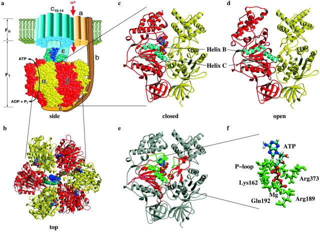
F0-F1 ATPase structure

There are three regions to which ATP is hydrogen bound. First, the so-called Walker or P-loop (residues β-Gly-159-β-Val-164) at the beginning of helix B. Second, the beginning region of helix C, namely β-Arg-189. And third, the residue α-Arg-373 from the α-subunit (see Fig. 1, c–e) (Abrahams et al., 1994). Sequential formation of these 15–20 hydrogen bonds ensures nearly constant force generation throughout the whole duration of the binding transition. This “binding zipper” sequence would lead to the smooth closing motion of the pocket and continuous conformational changes throughout the β-subunit (Oster and Wang, 2000a; Elston et al., 1998; Oster and Wang, 2000b).

In Results:
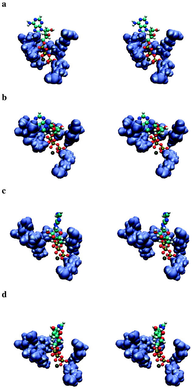
Stereo pictures of the hydrogen binding region and ATP. (a) Closed state, (b) 8th step, (c) 13th step, (d) open state. The orientation is the same as in Fig. 1 f.

Fig. 4, a–d provides four representative snapshots of the equilibrated closed and open binding pockets according to our simulations. These show that, in the closed pocket, the ATP molecule is surrounded by all three hydrogen binding regions. By contrast, in the open pocket a space has formed between the P-loop on one side and the helix C and the pocket's α-subunit region on the other side, with a gap between the ATP molecule and the P-loop so that ATP stays close to the α-subunit and helix C regions. An equilibrated intermediate structure where the pocket is half open is shown in Fig. 4 b. In that state the α-phosphate oxygen of ATP is still close to the P-loop, but the distance is already increasing between the β/γ-phosphate oxygens and the P-loop. The phosphate axis has rotated ∼30° and ATP is bridging the pocket. At the end of our simulations (Fig. 4 d), the ATP molecule is located between the two subunits as expected after its primary movement into the binding pocket. In that weak binding state, a newly docked ATP is expected to have contacted the ATPase, but not yet have induced conformational changes. Our simulations are consistent with this expectation. Further, we see that contacts are formed mainly between ATP and Mg2+, and between Mg2+ and the binding pocket.

In Discussion:

The importance of β-Lys-162, β-Arg-189, and α-Arg-373, for example, was connected to their formation of hydrogen bonds with ATP. In our simulations, these residues form the strongest and the last to break hydrogen bonds with the γ-phosphate oxygens. In addition, α-Arg-373 forms a hydrogen bond with one of the α-phosphate oxygens, acting as a restraining force for ATP during the movement of the P-loop and therefore facilitating the migration of the α-phosphate oxygens along the P-loop.

You can find further information here or here.

$\endgroup$
6
  • $\begingroup$ It's interesting that this source seemingly conflicts with the uniprot and interpro records from @JordiZambrino. I must say I can't find anywhere in that article that explicitly says it acts as a regulator specifically - the α-subunit still sounds to be intimately involved in catalysis. $\endgroup$
    – James
    Commented Apr 15, 2016 at 9:39
  • 1
    $\begingroup$ @James I too couldn't find explanation for alpha subunit's regulatory role. The article justs points out that it helps in stabilising ATP but doesn't say a word about regulation. But since wikipedia clearly states this, so I quoted it to them. Maybe the author of wikipedia article cited wrong article for that line or misinterpreted the conclusion. $\endgroup$ Commented Apr 15, 2016 at 9:50
  • $\begingroup$ Why the downvote??? Please tell me so that I could improve any mistake in the post. $\endgroup$ Commented Apr 15, 2016 at 14:02
  • $\begingroup$ Would you agree that the article you have linked to is basically saying that the $\alpha$ subunit's role is to increase the efficiency of ATP-ase by allowing more locations for hydrogen bonds to form with? (btw the downvote wasn't me :) ) $\endgroup$ Commented Apr 16, 2016 at 13:10
  • 1
    $\begingroup$ @Quantumspaghettification It seems so from what it (article) says, but it seems to me more that α subunit is for stabilizing ATP inside ATPase. I am continuously searching more details on this. $\endgroup$ Commented Apr 16, 2016 at 13:54
1
$\begingroup$

From: Uniprot

Mitochondrial membrane ATP synthase (F1F0 ATP synthase or Complex V) produces ATP from ADP in the presence of a proton gradient across the membrane which is generated by electron transport complexes of the respiratory chain. F-type ATPases consist of two structural domains, F1 - containing the extramembranous catalytic core, and F0 - containing the membrane proton channel, linked together by a central stalk and a peripheral stalk. During catalysis, ATP synthesis in the catalytic domain of F1 is coupled via a rotary mechanism of the central stalk subunits to proton translocation. Subunits alpha and beta form the catalytic core in F1. Rotation of the central stalk against the surrounding alpha3beta3 subunits leads to hydrolysis of ATP in three separate catalytic sites on the beta subunits. Subunit alpha does not bear the catalytic high-affinity ATP-binding sites.

Names and Taxonomy

From: InterPro

There is a substrate-binding site on each of the alpha and beta subunits, those on the beta subunits being catalytic, while those on the alpha subunits are regulatory. The alpha-subunit contains a highly conserved adenine-specific non-catalytic nucleotide-binding domain, with a conserved amino acid sequence of Gly-X-X-X-X-Gly-Lys. The alpha and beta subunits form a cylinder that is attached to the central stalk. The alpha/beta subunits undergo a sequence of conformational changes leading to the formation of ATP from ADP, which are induced by the rotation of the gamma subunit, itself is driven by the movement of protons through the F0 complex C subunit [PMID: 12745923].

Sources:

  1. http://www.ebi.ac.uk/interpro/entry/IPR005294
  2. http://www.uniprot.org/uniprot/P25705
  3. http://www.ncbi.nlm.nih.gov/pmc/articles/PMC3278611/
  4. http://www.ncbi.nlm.nih.gov/pmc/articles/PMC1679692/
$\endgroup$
0
$\begingroup$

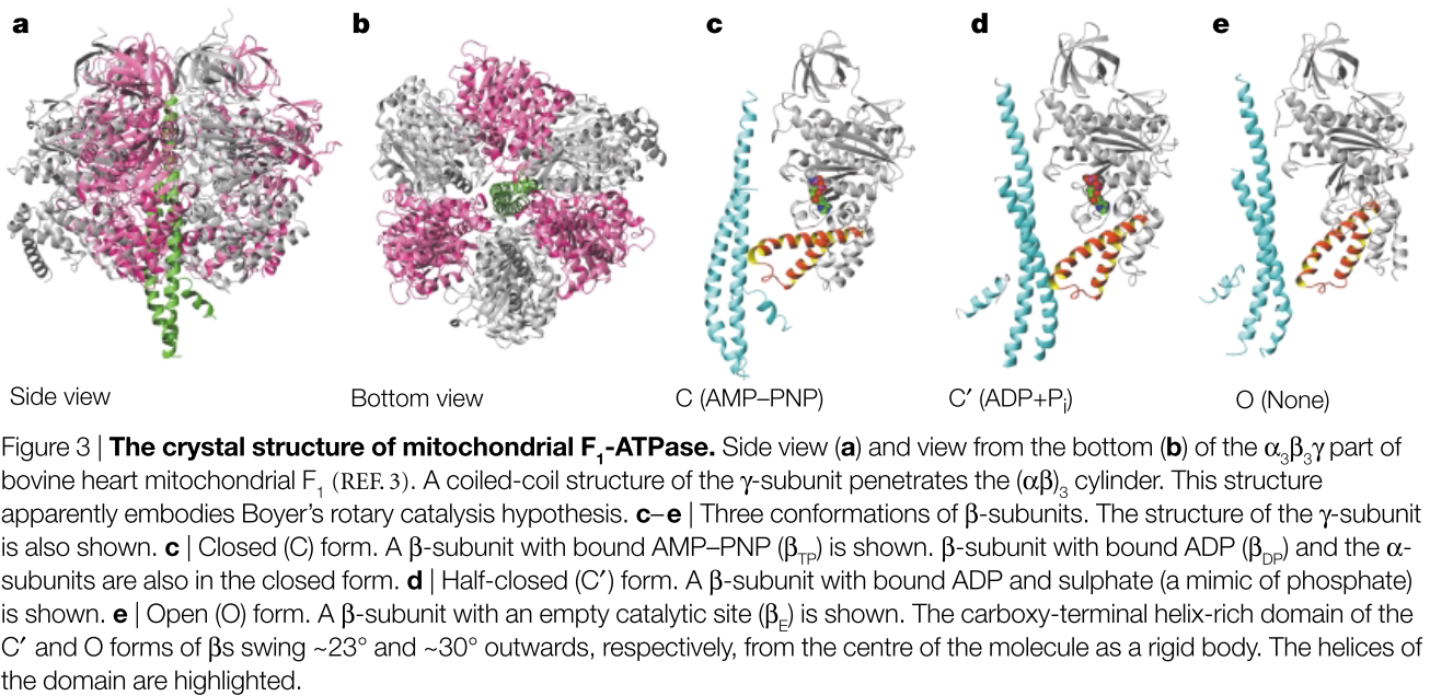
The α- and β-subunits have a similar fold, as would be expected from their sequence similarity. All of the α- subunits are bound to the ATP analogue AMP–PNP, and the three subunits adopt very similar conformations. The three β-subunits, however, are in three nucleotide-bound states: the first, termed βTP, has AMP–PNP in the catalytic site (FIG. 3c); the second (βDP) has ADP; and the third (βE) has no bound nucleotide (FIG. 3c–e). So, the native structure of F1 looks like a snapshot of the working rotary engine, with three reaction chambers representing the moment just after exhaust and intake (βΕ), ignition (βDP) and compression (βTP) (BOX 2). The lower part of the slightly bowing, asymmetric coiled-coil structure of the γ-subunit is displaced towards the βE , forcing the carboxy-terminal domain of this β-subunit to swing ~30° downwards. Thus, the βE adopts the ‘open’ (O) form, whereas βTP and βDP have the ‘closed’ (C) form.

enter image description here

Yoshida, M., Muneyuki, E., & Hisabori, T. (2001). ATP synthase--a marvellous rotary engine of the cell. Nature Reviews. Molecular Cell Biology, 2(9), 669–77. http://doi.org/10.1038/35089509

$\endgroup$

You must log in to answer this question.

Not the answer you're looking for? Browse other questions tagged .