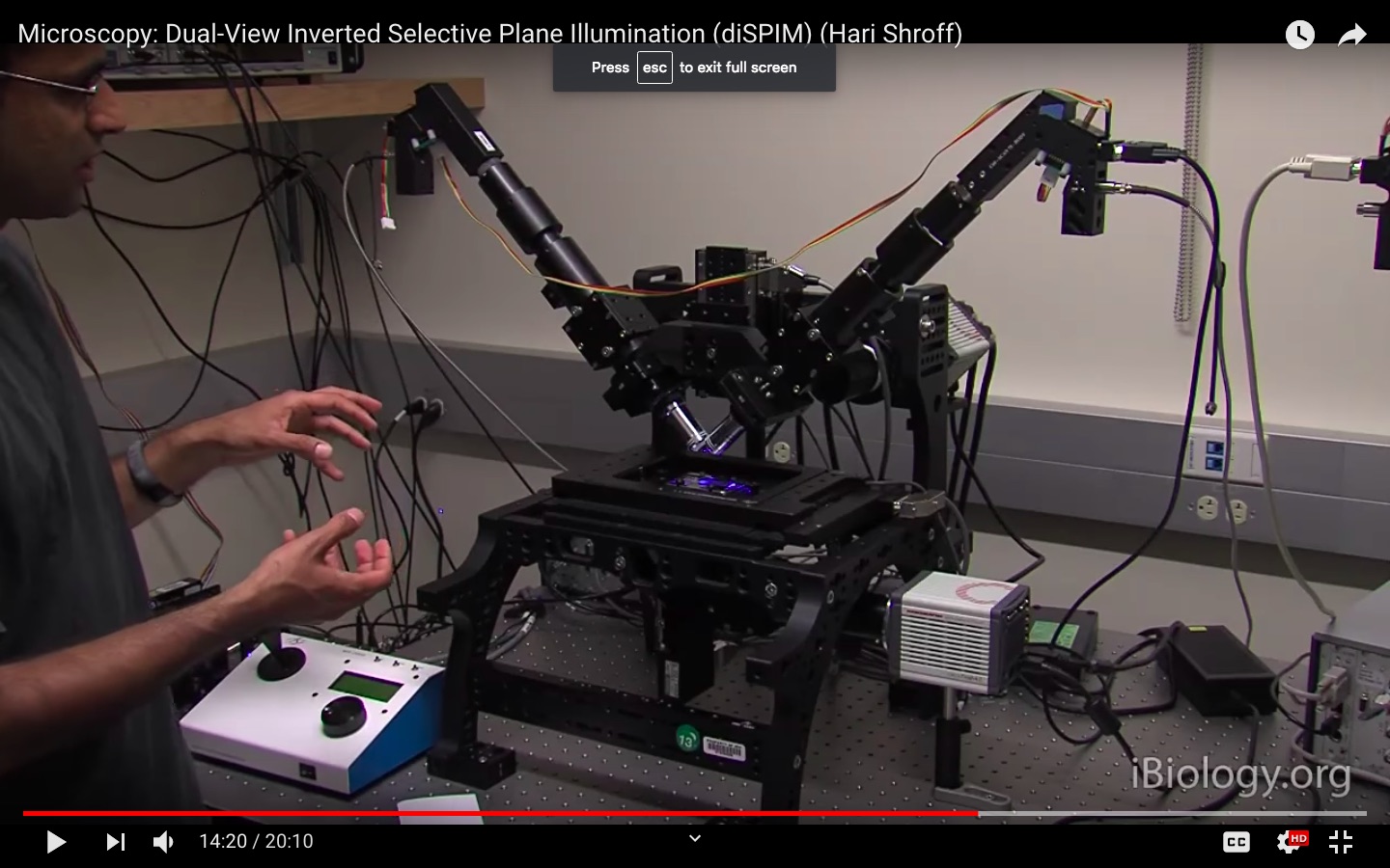
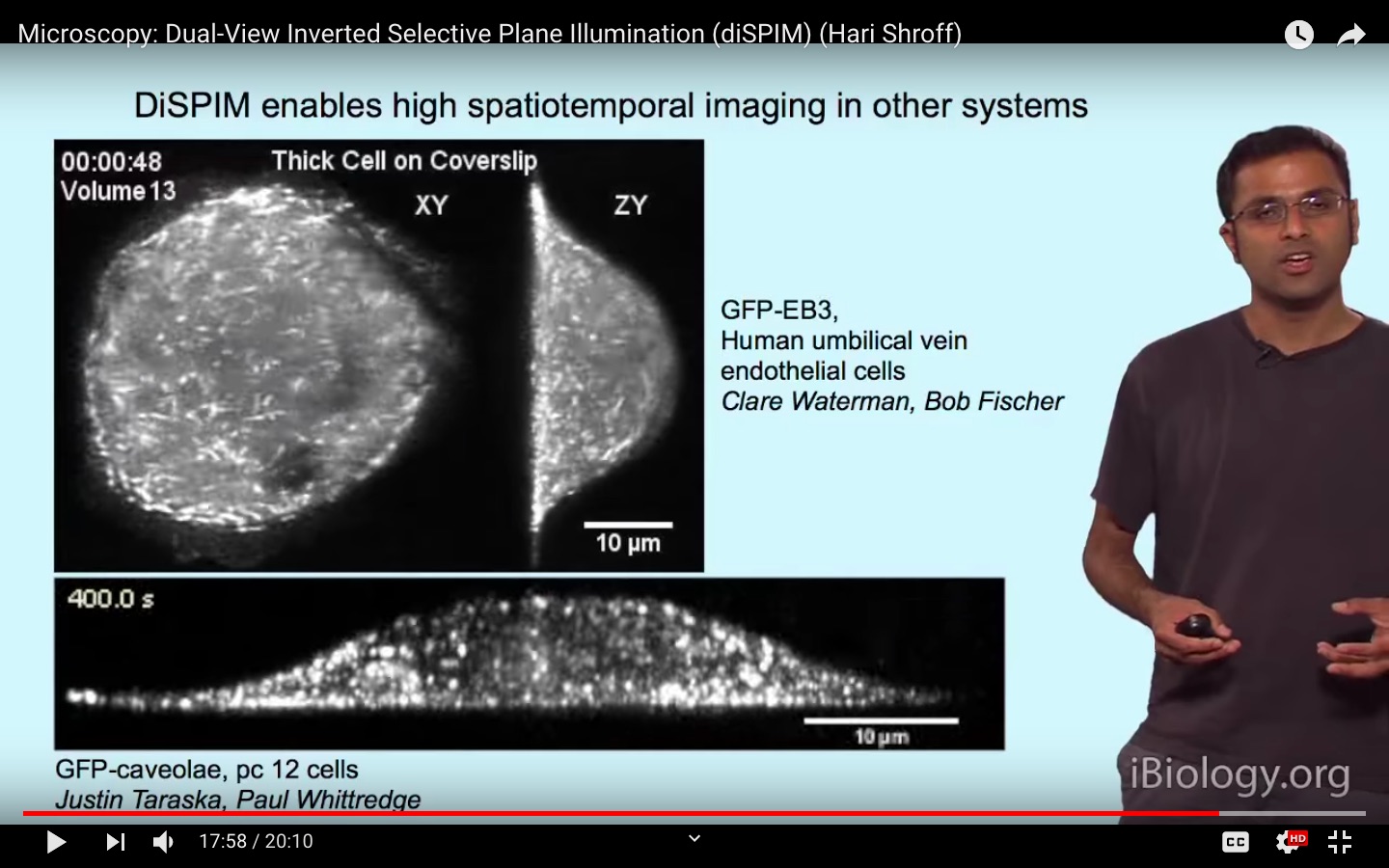
The iBiology Techniques video Microscopy: Dual-View Inverted Selective Plane Illumination (diSPIM) (Hari Shroff) describes light sheet microscopy and the improvement in resolution by introducing dual-view (two cameras) capability.
What's happening is that the object is moving randomly in 3D, and so for each frame, the stacks are used to generate a 3D image, and then it is de-rotated to synthesize a view in a plane fixed to the object, rather than the microscope.
Is there a simple way to understand how these image stacks are processed to obtain a smooth 3D reconstruction that can then be sectioned computationally? Is there some kind of interpolation or modeling of structure at the limit of the depth resolution?
Rather than a complete explanation, which would likely be fairly mathematical and in depth, a simpler explanation plus a reference or link to a more in-depth description might be a better way to help me get on the right track.


