3
$\begingroup$

I am learning about the endocochlear potential. According to different sources, the ionic composition of perilymph is about as follows (in mM): 150 Na+, 4-5 K+, 1.2 Ca2+, 1 Mg2+, 120 Cl−, and 20 HCO3−; and that of endolymph is 1 Na+, 157 K+, 0.02 Ca2+, 0.01 Mg2+, 132 Cl−, and 30 HCO3−.

This means (in a naive attempt to quantify ionic charges) that the sum of charges in perilymph is 150+4.5+2*1.2+2*1-120-20 = +18,9. And in endolymph it is: 1 + 157 + 2*0.02 + 2*0.01 - 132 - 30 = -3,94.

From these numbers, it seems like perilymph contains more positive charges than endolymph. How is it possible that the potential of endolymph is highly positive (80-90 mV) compared to perilymph? Shouldn't it be the other way around?

I have searched the forum and the internet for answers, but my question seems so basic (or based on a very fundamental misunderstanding) that I can't find anything of help. The only explanation I keep seeing is that the high potassium concentration accounts for the potential. However, isn't that outweighed electrically by the excess of negative ions in contrast to the perilymph?

$\endgroup$
8
  • $\begingroup$ Not an idiot or stupid at all, please don't describe yourself that way - in fact since it is not true I will edit it out of your question :) You have a very common misconception for people studying this material, which is not realizing that the positive and negative concentration inside and out is effectively equal (you just don't have the full list). See if this previous answer: biology.stackexchange.com/questions/79619/… helps out. If not, I can tailor a fresh answer here. $\endgroup$
    – Bryan Krause
    Commented Aug 28, 2019 at 15:32
  • $\begingroup$ Hello Bryan! Thank you so much for your nice reply! Unfortunately, I still don't quite understand it. As a matter of fact, I think I may be even more confused, and I'm not sure if I can put into words what exactly why. I will give it a try. First, if the charge concentrations are the same on both sides, shouldn’t the measurable voltage be zero? A potential of 90mV designates that an electric (not chemical) potential is actually measurable across a membrane, doesn’t it? Why is that if the electric net is the same of both sides? $\endgroup$
    – Jimmy
    Commented Aug 28, 2019 at 17:24
  • $\begingroup$ My simplified understanding of electrochemical potentials used to be like this: if there is a concentration gradient across a membrane (for example of KCl), which is only permeable for K+ (but not Cl-), K+ will stream to the side of lower [KCl] to reach equal distribution. However, as no equal amount of negative charges follow (!), the charge concentrations will be different on either side and due to repulsion/attraction, an equilibrium between ‘chemical driving force’ and ‘electric driving force’ will be reached at some point. $\endgroup$
    – Jimmy
    Commented Aug 28, 2019 at 17:25
  • $\begingroup$ If the charge concentrations remain basically the same on both sides, as you just said, how does the electric component exist at all? Thank you!! $\endgroup$
    – Jimmy
    Commented Aug 28, 2019 at 17:25
  • 1
    $\begingroup$ Thank you a 1000 times in advance. I'm looking forward to your answer! $\endgroup$
    – Jimmy
    Commented Aug 28, 2019 at 19:16

1 Answer 1

1
$\begingroup$

About biological electrical potentials generally

Biological potentials on the order of tens of millivolts depend on very very few ions moving (about 1/100,000 of the potassium concentration, for example, for a typical neuronal potential).

Therefore, the sum of positive and negative charges in any compartment is always almost zero (to several decimal places and beyond measurement; if you say a ). Electrical forces are very powerful, and

Some previous answers that are related:

https://biology.stackexchange.com/a/57066/27148

https://biology.stackexchange.com/a/77925/27148

https://biology.stackexchange.com/a/81571/27148

The endocochlear potential in particular - this is really cool

Everything I said above basically can be summarized in a quick statement: you can't figure out potentials just by measuring ion concentrations and summing them up. If you counted all the ions (including charged proteins, etc), you'd get ~0 in any biological solution. Instead, you have to think about where the concentration gradients are and how particular ions are moving. Let's tackle the endocochlear potential... Hibino et al 2010 has an excellent diagram, adapted from their previous 2008 paper:
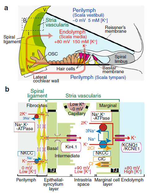
Structure of the cochlea and its lateral wall, from Hibino et al 2010

Let's walk through the key features here, focusing on panel (b). First, the general structure: there are three extracellular spaces, which are all electrically isolated from each other. Between these extracellular spaces, there are two layers of cells where all the business happens.

Between the perilymph and intrastria space, there are a collection of cell types all connected via gap junctions. We can treat them sort of like one big cell, I'll call it the syncytium. A sodium-potassium ATPase uses energy to pump potassium into the syncytium and sodium out (upper left of [b]). Additionally, because there is now low sodium in the syncytium, more potassium enters through NKCC channels, which are symporters that move 1 sodium, 1 potassium, and 2 chloride ions together.

Potassium would accumulate in the syncytium, except there are Kir4.1 channels permeable to potassium on the other side of these cells, open to the intrastria space. So, potassium flows down its concentration gradient into the intrastria space. This also creates an electrical potential and makes the intrastria space very positively charged (shown as +90mV on the diagram; the syncytium would similarly get a negative charge from the flowing potassium, but that isn't shown).

Normally, not many ions would actually flow to create this potential (on the order of the 1/100,000 I mentioned above) and everything would stop there. However, there are another set of pumps and channels in the marginal cells. The marginal cells also pump potassium in, via the sodium-potassium ATPase and NKCC channels, keeping potassium concentrations in the intrastria space low, such that potassium continues to flow down its concentration gradient from the syncytium.

The marginal cells also have potassium channels on their opposite face, and these are open to the endolymph. The high concentration of potassium ions in the marginal cells and the positive charge in the intrastria space gives the necessary drive to push the concentration of potassium in the endolymph high.

Ultimately, there is a full circuit of potassium because potassium is flowing into hair cells from the endolymph, and then back out to the perilymph. The sodium-potassium pumps use energy in the form of ATP to maintain the imbalance between perilymph and endolymph.


Hibino, H., Nin, F., Tsuzuki, C., & Kurachi, Y. (2010). How is the highly positive endocochlear potential formed? The specific architecture of the stria vascularis and the roles of the ion-transport apparatus. Pflügers Archiv-European Journal of Physiology, 459(4), 521-533.

Nin, F., Hibino, H., Doi, K., Suzuki, T., Hisa, Y., & Kurachi, Y. (2008). The endocochlear potential depends on two K+ diffusion potentials and an electrical barrier in the stria vascularis of the inner ear. Proceedings of the National Academy of Sciences, 105(5), 1751-1756.

$\endgroup$
4
  • $\begingroup$ Hello Bryan, thank you so much for your explanation. I think I got a better idea now. There are just two things I'm still confused about. 1) You say that the sum of all charges is 0 in any biological context. Why do electrodes on either side of the membrane measure +90mV then? There must be more + ions that - ions on the more positive side for that to happen, or do I misunderstand the entire concept of voltage? 2) The K+ flowing into the intrastria space cause a positive potential. That made sense to me until I read they are effectively removed. Shouldn't the potential leave with them? $\endgroup$
    – Jimmy
    Commented Sep 1, 2019 at 13:58
  • $\begingroup$ To clarify, I do understand the flow directions and the dependences on concentration gradients. It is the voltage measurements that throw me off. I don't understand how an electric potential is reconcilable with the same net charges on both sides, and why the voltage between syncytium and intrastrial space would still be positive after the positive ions moved on already. $\endgroup$
    – Jimmy
    Commented Sep 1, 2019 at 14:02
  • $\begingroup$ @Jimmy Check out the first other answer I linked, I have a couple back of envelope calculations for how many ions are responsible for a biological potential. It works out to be something like 1/100,000. So although the concentration of positive to negative ions is equal out to at least 3-4 digits (well beyond any ability to measure actual concentration) it's that tiny little bit that makes the difference. $\endgroup$
    – Bryan Krause
    Commented Sep 1, 2019 at 14:59
  • $\begingroup$ For the intrastrial space, potassium is getting pumped out but other ions are moving too between the marginal cells. Probably the better way to think about the +90mV is that if the space is less than +90mV, more potassium would flow from the syncytium and the potential would rise. +90mV is the voltage at which the net flow from syncytium to intrastrial equals the net flow from intrastrial to marginal. (same for the endocochlear potential itself) $\endgroup$
    – Bryan Krause
    Commented Sep 1, 2019 at 15:04

You must log in to answer this question.

Not the answer you're looking for? Browse other questions tagged .