The scientific question
The poster is interested in how the fluorescent protein, aequorin, catalyses the production of light. He mentions the requirement for calcium and oxygen, the role of coelenterazine as a “luciferin”, and is aware of the fact that the protein undergoes an conformational change. He specifically asks
“what part of the protein structure [my emphasis] actually catalyses
this reaction after the conformational change”
I think this is best answered — and will be more useful to those who are not familiar with this protein — by first considering the overall process, and then looking at what is known about changes in structure and how this can be interpreted.
The sequence of event in aequorin fluorescence
Fluorescence of aequorins such as aequorin or obelin occurs through a cycle of events:
- The protein (apoprotein) non-covalently binds the organic prosthetic group, coelenterazine, and molecular oxygen
- Coelenterazine is ‘activated’ by oxidation to 2-hydroperoxycoelenterazine†
- Calcium ions bind to the apoprotein
- Activated coelenterazine is converted to coelenteramide and molecular carbon dioxide with concomitant release of blue light
- Coelenteramide and calcium ions are released from the protein
I would imagine that coelenteramide would be converted back to coelenterazine by some process, rather than being degraded, but I have not been able to find information on this point.
†In the papers I looked at for this answer I did not encounter information on this, albeit important, aspect of the topic. No doubt such information exists.
The substrate and effector binding sites on the protein
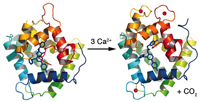
Our knowledge of the molecular details of catalysis by proteins depends on determining structures in which the protein is ‘frozen’ at one particular stage of a reaction. In this case it is not too difficult to crystalize the protein bound to either coelenterazine or to calcium ions, but the reaction and release of these molecules hinders finding a stable structure with both bound. The structures below from the work of Liu et al. show obelin, first with bound coelenterazine, and then with bound calcium ions, although here the coelenterazine has been converted to coelenteramide. This is useful for seeing the relative positions of ions and prosthetic group within the apoprotein.

It can be seen that the three calcium ions (red balls) are bound to similar helix–loop–helix domains called EF hands which are also found in other calcium-binding proteins. In fact, the molecule has pseudo-symmetry with four such domains, but one of them does not bind calcium. The large binding site for coelenterazine is in the centre of the molecule and involves parts of the helices of the EF hands. Because of the cartoon representation, which ignores the side-chains, the binding site appears much more open than is the case.
Conformation changes on binding calcium ions
Let us consider this first from a general standpoint. Because calcium ions bind to the amino acid side-chains of EF-hands through many polar interactions and hydrogen bonds, there is a structural difference between the bound state and the unbound state, where different interactions of these side-chains will occur. Such structural changes at the calcium-binding sites are accompanied by structural changes at distal positions in the protein, including the site at which a substrate binds. This, in turn, can affect the conformation of the substrate, facilitating its reaction. Although it may be most appealing to consider the effect of the binding of calcium or other effectors as initiating a concerted chain of events, it is perhaps more often regarded as affecting the position of equilibrium between two alternative conformations of a protein, differing only slightly in thermodynamic Gibbs free energy. This latter relates more easily to the comparison of two crystal structures of a protein, where there are “before” and “after” situations to compare.
In the case of obelin, the two structures (above) show anticipated, although relatively modest, structural differences at the calcium binding site. I do not reproduce them here as they are best inspected with a pair of stereo glasses in a printed copy of the paper Liu et al.. The same is true for the changes in the substrate binding site, but it can be seen from the figure above that there is a change in conformation of the substrate after carbon dioxide is released.
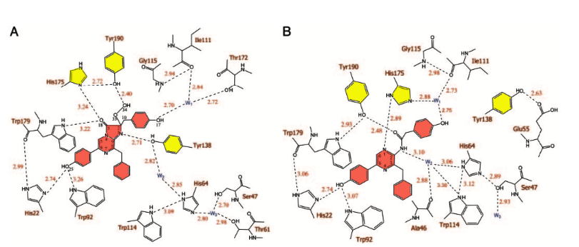
The two-dimensional comparison (below) is pertinent as it shows (as does the work of others) that there is a particularly marked change in the interaction of a specific tyrosine and histidine residue (Tyr190 and His175 in obelin, here, and Tyr184 and His169 in aequorin and that of Tyr138 (Tyr132 in aequorin) at the other ‘side’. Changes in bonding to water molecules are also seen (discussed below).

[From Liu et al. (2006) Comparison of the hydrogen-bond network in the binding cavities of obelin before (A) and after (B) discharge of calcium ions.]
How does the conformational change cause bioluminescence?
The mechanism of bioluminescence is outside my sphere of competence, and ideas on how this is triggered in aequorin are based on a range of other experiments. However the following may be of interest:
In 2000 Head et al. wrote, regarding aequorin:
Displacement of the helices flanking the C-terminal loop would be
expected to disrupt the hydrogen bonding of the loop, resulting in
relocation of the side chain of Tyr 184 [Tyr190, above]. This is in
turn would disrupt the hydrogen bonds to His 169 [His 175, above] and
the peroxide. No longer stabilized, the peroxide would be free to
attack the carbonylic C3 to initiate the light-emitting reaction.
Liu et al., writing in 2006, elaborated on this by including a role for the water molecule (W2), the change of position of which they had observed (above). They proposed the following reaction scheme:

How does a physical scientist go about finding — and understanding — information on proteins?
In my original answer I focussed on how to find information about proteins. That I retain in a more condensed form below. It appears that the poster has a strong background in the chemistry relating to this work, but has had little contact with proteins. As someone whose first degree was in chemistry and then moved into molecular biology, I sympathize. However, although one’s chemistry makes it easy to understand the chemical principles in protein chemistry, one still has to become familiar with the structure of proteins and accustom oneself to what may seem a strange world of weak interactions and non-standard environments. There is no avoiding the need to read at least the chapters on proteins and enzymes in a solid text like Biochemistry by Berg et al., and for this purpose any old second-hand edition will do.
With this background one can, of course, use a general search engine or Google Scholar to search for information on a particular protein. However when these methods fall short I usually go to the Protein Data Bank and search for the protein involved. In this case I used the two terms, aequorin and coelenterazine. This gave me a choice of different structures. I browsed through them, had a look at the 3D views, and then tried to find a relatively recent structure which had a publication associated with it. In this case I chose 7O3U.pdb, which has a related 2022 paper in Protein Science that seemed to discuss the mechanism and active site. From there I worked back to the two papers which are cited in this answer.


