On Wikipedia it is explained that the human eye has a certain 'resolution'. Does this mean that the retinal image consists of pixels? If yes, what shape would these pixels have?
-
1$\begingroup$ Are you asking about the shape of the actual physical representation (rods/cones) or the subjective visual impression? $\endgroup$– fileunderwaterCommented Jan 19, 2015 at 15:57
-
$\begingroup$ I can't imagine this meaning anything other than physical representation, since we do not actually perceive pixels at all. $\endgroup$– March HoCommented Jan 19, 2015 at 18:23
-
$\begingroup$ What shape are stars? Presumably a point light source would illuminate precisely one 'pixel' and you would perceive the shape of that pixel only. In terms of the actual cells, they're roughly round when looked at from 'above'. They're round tubes. Like straws. $\endgroup$– ResonatingCommented Jan 19, 2015 at 22:25
-
1$\begingroup$ Related: What's the smallest size a human eye can see? $\endgroup$– fileunderwaterCommented Jan 19, 2015 at 23:03
-
2$\begingroup$ You need to realize that the human eye doesn't in any way work as a camera. Only a small area of our eye can see sharp images at high resolution, and resolution and sharpness also differs over the rest of the retina as well. So the resolution varies over the retina. The image we actually see is also a continiously interpreted and updated, and the brain is very creative in this process by basically "filling in the blanks". We therefore don't see in pixels. $\endgroup$– fileunderwaterCommented Jan 19, 2015 at 23:18
2 Answers
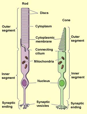
The pixels of the visual system are represented by the photoreceptors. Their shape is the following (adapted from "The Brain from Top to Bottom, MgGill University"):

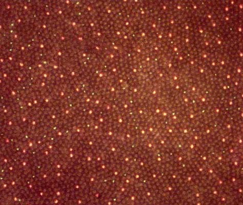
There are about a 180 photoreceptors packed on 288 microns on the retina (see "What's the smallest size a human eye can see?") so about 1 pixel per 2 microns. The rods and cones are packed closely together on the retina, referred to as the retinal mosaic. This retinal mosaic is not unlike a photosensitive chip, as can be seen on the folllowing image (Developmetal Neurobiology lab, UCLA, Santa Barbara):

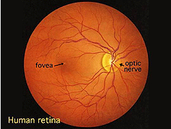
We do not visually perceive this mosaic, as the optics of the eye (e.g., light diffraction by the cornea and the lens, and distortions due to the relatively large pupil size) prevents optimal use of the photoreceptor density (the maximal resolution). Furthermore, especially outside the field of view (the foveal region), the output of many photoreceptors are funneled onto one single optic nerve fiber, thereby fusing the output of many photoreceptors (See the question "Why can we not see around our point of focus of our eyes?"). Lastly, the brain actively 'glues' any gaps in the visual field together. Most strikingly, the blind spot, which is the place where the optic nerve exits the eye is fairly large, is normally not visible to us: see the following picture (PNAS online content):

Hence, any gaps in the retinal image are in fact actively smoothened out by the brain.
-
$\begingroup$ Interesting answer. In the field of view do the photoreceptors each connect to a single nerve? Roughly speaking how many photoreceptors may channel into a single nerve fiber, on average (unless there are actual values for different areas of the eye!)? This would ultimately be the smallest "resolution" or "pixel size" then, rather than the size of the photoreceptor itself. Thanks again $\endgroup$– LukeCommented Jan 21, 2015 at 13:55
-
$\begingroup$ Thanks. Good question :) As to neural retinal connectivity see: "Why can we not see around our point of focus of our eyes?" You can click the link in the answer above. $\endgroup$– AliceD ♦Commented Jan 21, 2015 at 13:58
If you by pixels are referring to the photoreceptors (rods & cones), their density differs over the retina, and we can only see sharp images at high resolution in a small region of our eye (fovea). So the resolution varies over the retina. The image we actually see is also a continiously interpreted and updated, and the brain is very creative in this process by basically "filling in the blanks". We therefore don't see in pixels, and our resolution is influenced by (at least) light conditions, contrast, photoreceptor density, signal merging and interpretation of the visual signals by the visual cortex in the brain. However, our eye is also constantly scanning the visual field (see saccade), so even if the "static" visual resolution (when we focus constantly on a single point) is drastically different over the visual field, this does not match our visual perception. As a sidenote, saccades are possibly the fastest human motions, and can reach speeds well above 400°/s (Boghen et al, 1975) - wikipedia reports 900°/s but without a clear reference - and takes place several times per second.
If we focus at the resolution in terms of photoreceptor density (densities of rods/cones), this is drastically different over regions of the retina. For instance, Jonas et al. (1992) reports ~150,000 cones/mm2 close to the center of the fovea to 2500-6000 cones/mm2 towards the periphery. The approximate density of rods goes from 150,000 rods/mm2 to 30,000 in the periphery. Cone & rod diameter also differs over the retina, with larger diameters towards the retinal periphery, which relates to light sensitivity.
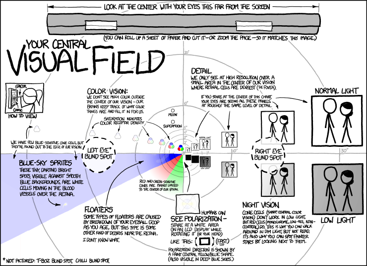
A nice summary of the flexibility and limitations of human vision can be seen in this xkcd comic (click for larger version):
 (from "xkcd: Our central visual field" at http://xkcd.com/1080/)
