2
$\begingroup$

I had read in a paper that present a low noise amplifier the following:

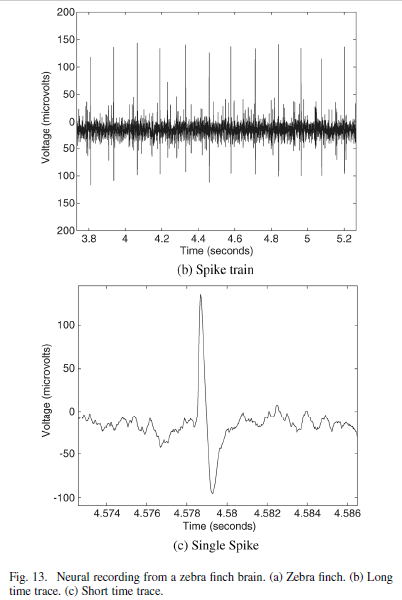
"...This level of input signal is larger than both typical action potentials (<500μV) and local field potentials (<5 mV). If a larger input signal is expected, the amplifier’s supply voltage can be increased at the expense of higher power consumption.We verified that our neural amplifier works in a real recording environment by using it to record action potentials in the robust nucleus of the arcopallium (RA) of the anesthesized zebra finch. Data were taken with a carbon fiber electrode (Kation Scientific, Inc.) electrode that had an impedance of approximately 800kHz at 1 KHz. A long extracellular trace and a short extracellular trace recorded from our amplifier (normalized by its gain) are shown in Figure 2. They were found to be identical to that recorded by a commercial neural amplifier (A-M Systems, Model 1800)."

Link:

http://www.rle.mit.edu/acbs/pdfpublications/journal_papers/woradorn_tbcas_neural_amp.pdf

Please, can someone help me to understand how we can measure an action potential as shown in figure 2? We know that action potential amplitude is approximately 70mV as shown in figure 1 but figure 2 shows that is approximately 70μV.Is this possible?

Figure 1

enter image description here

Figure 2

enter image description here

$\endgroup$

1 Answer 1

1
$\begingroup$

I'm going to mostly duplicate my answer to another question Neural signals amplitude since it's exactly the same as this one, but it has no upvotes so I can't vote as duplicate. Once I get an upvote either place I'll vote to close the other.

In the first figure, those voltages refer to the difference between the inside and outside of the cell (i.e., -70mV means the cell is 70mV more negative than the outside).

You can't possibly measure that potential difference from outside of a cell. When you do extracellular recording, like in the second figure, you are measuring spatial differences in ion concentrations that occur due to ions flowing in and out of cells. For example, when a cell fires an action potential, lots of positive ions flow into the cell; this makes the area just around the cell slightly more negative than an area further away. The magnitude of this potential as measured depends greatly on your recording configuration, the type of signal you are recording (i.e., population activity or just a single cell; synaptic currents versus action potentials), how close your electrode is to the signal you are recording, and where your ground is (or position of both of your electrodes if you are doing a bipolar recording).

Also, in this second recording, either the voltage is reversed (i.e., the electrode near the cell is taken as ground), or it is actually measuring return currents in the dendrites of the cell; otherwise, the action potential would be in the opposite polarity (downward first, then upward) compared to inside the cell.

Although it varies, in most cases the signals you record extracellularly are on the order of microvolts to a couple of volts.

Lastly, in the quote you reference, the line before it was:

The total harmonic distortion (THD) of our amplifier stays below 1% for input signals smaller than 7.3 mV peak-to-peak.

That article is describing an amplifier system for recording extracellularly (potentially for a brain-machine interface). They are saying that one type of distorting noise is not a problem in their amplifier at the input voltages you would expect in an extracellular recording of spikes or local field potentials, which indeed would usually be less than 5 mV. This amplifier would not be suitable for recording intracellularly.

$\endgroup$
4
  • $\begingroup$ Thank you so much mr Bryan Krause for your response. Sorry for my comment but I would to understand one point.You refer that "That article is describing an amplifier system for recording extracellularly "and also "This amplifier would not be suitable for recording intracellularly."Can you help me to understand the difference when amplifier records intracellularly compared with extracellularly ?What changes in neural signals that record? Maybe the difference is the place of implant in brain?Is any of the recording methods better? Thank you in advance. $\endgroup$
    – elecV1
    Commented Aug 21, 2017 at 19:14
  • 1
    $\begingroup$ Read about whole-cell patch clamp (though there are a couple other methods for recording intracellularly). Intracellular recordings are necessary to measure the magnitude of inputs to individual cells. Extracellular recordings can measure outputs from single or multiple cells, and/or field potentials that approximate inputs to populations of cells. It isn't fair to say one is better than the other: they are different, for different purposes. $\endgroup$
    – Bryan Krause
    Commented Aug 21, 2017 at 19:20
  • 1
    $\begingroup$ For long-term recordings (over weeks and months), only extracellular recordings are currently technologically feasible. $\endgroup$
    – Bryan Krause
    Commented Aug 21, 2017 at 19:21
  • $\begingroup$ Thank you so much, Mr Bryan Krause. I search for this answer for days! $\endgroup$
    – elecV1
    Commented Aug 21, 2017 at 19:32

You must log in to answer this question.

Not the answer you're looking for? Browse other questions tagged .