3
$\begingroup$

Modern electronic sound recording equipment employs a physical membrane that triggers the piezoelectric effect in a metallic element, to transform sound waves into electric signals.

I had always thought that the eardrum (or tympanic membrane) was the main instrument of hearing in vertebrates, that it physically transduces sound waves into nerve signals. However, in looking into it, I find that the eardrum transmits sounds to inner ear anatomy, where more structures are encountered before the sound waves become nerve signals.

I also find that small hairs, cilia, are sensitive to sound, and appear to be what actually turns sound waves into nerve signals, perhaps analogously to rod and cone cells in the eye. These cells line the ear canal, and when I read about them, they seem to be the real mechanism of hearing, and not the ear drum or inner ear bones.

At this point I'm confused as to actually how sound waves are transduced into nerve signals in vertebrates. Can someone explain the the roles of the various parts of anatomy in vertebrate hearing, in the overall, big picture? What roles do the large parts play, versus the microscopic cilia?

$\endgroup$

2 Answers 2

2
$\begingroup$

Sounds are pressure waves in air, but the inner ear is a liquid-filled space. This presents an impedance matching problem where sound is reflected rather than transmitted.

The eardrum and inner ear bones perform this mechanical impedance matching/transduction of air-to-liquid sound. From Purves' Neuroscience:

Sounds impinging on the external ear are airborne; however, the environment within the inner ear, where the sound-induced vibrations are converted to neural impulses, is aqueous. The major function of the middle ear is to match relatively low-impedance airborne sounds to the higher-impedance fluid of the inner ear. The term “impedance” in this context describes a medium's resistance to movement. Normally, when sound travels from a low-impedance medium like air to a much higher-impedance medium like water, almost all (more than 99.9%) of the acoustical energy is reflected. The middle ear (see Figure 13.3) overcomes this problem and ensures transmission of the sound energy across the air-fluid boundary by boosting the pressure measured at the tympanic membrane almost 200-fold by the time it reaches the inner ear.

Some sources will refer to this as "amplification" which is correct in some ways (without it, sound would be too "weak" in the inner ear) but doesn't quite explain the whole problem.

Inner hair cells do the actual sensory transduction, converting vibrations into electrical signals when the "hairs" are stretched apart mechanically, causing a fragile "tip link" to pull on a physical channel, opening it and allowing ions to flow through. The cochlea is a frequency-analyzer, vibrating at different frequencies along its length and allowing different hair cells to respond maximally to different frequencies of sound.


As a side note, although you asked about vertebrates, aquatic vertebrates like fish don't have these middle ear bones, their hearing is quite different from the mammalian ears I am most familiar with. I'm not sure at all about hearing mechanisms in aquatic mammals that evolved from terrestrial ancestors, like whales and dolphins, but that might be a good subject of a later question.

$\endgroup$
0
$\begingroup$

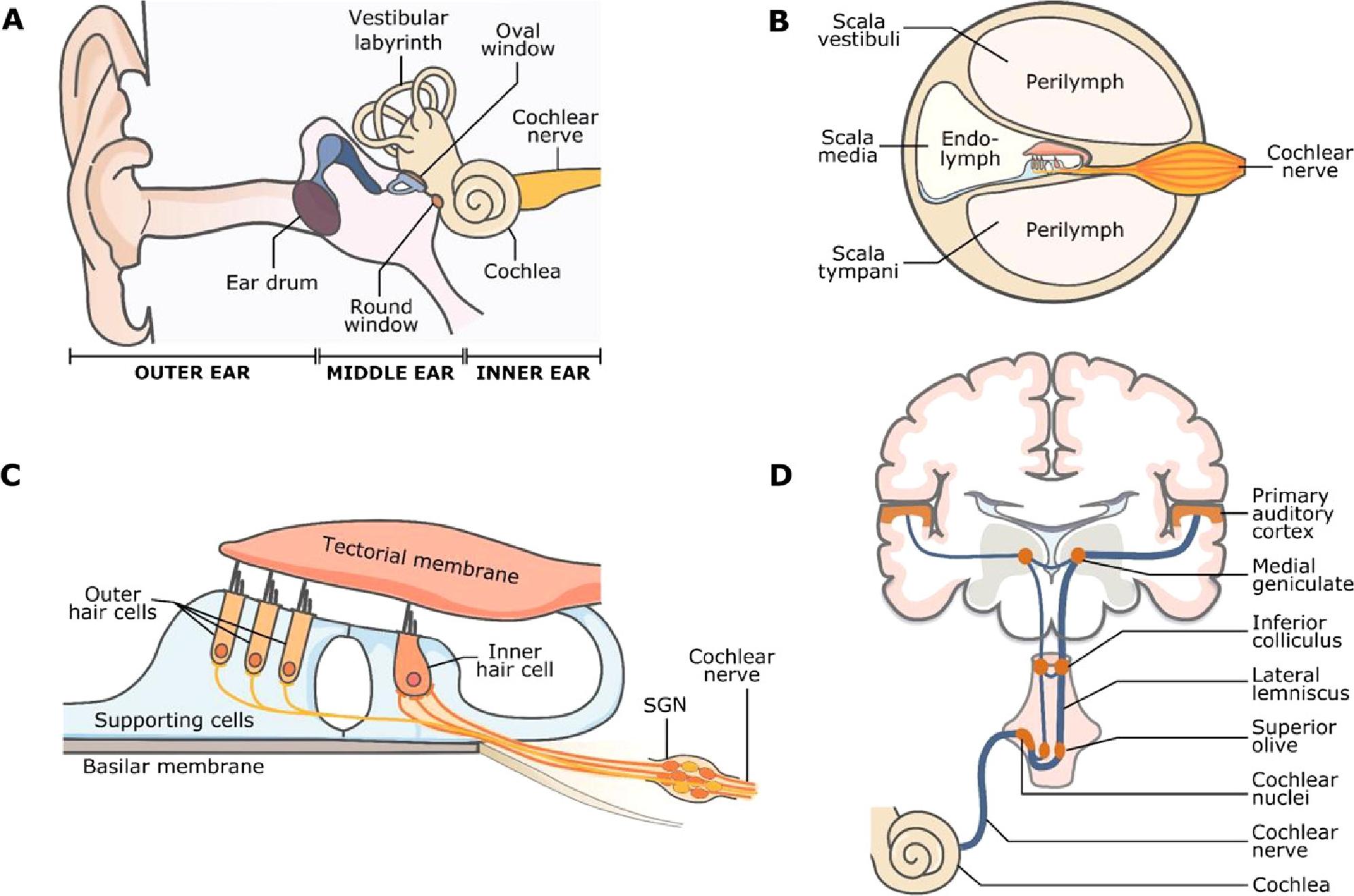
The auditory system (Fig. 1) is basically comprised of 3 main parts - the outer, middle and inner ear (the cochlea).

Generally spoken, the outer ear captures sounds, the middle ear transmits them, the inner ear acts as a transduction system to translate the acoustic pressure waves into electrical signals.

  • The pinna of the outer ear funnels acoustic waves into the ear canal (meautus) and amplifies it. The pinna changes the transfer function that aids in sound localization in the vertical plane (Fig. 1A).
  • The middle ear conveys the pressure waves from the ear drum (tympanic membrane) to the ossicle chain, amplifying the sound further (Fig. 1A).
  • The inner ear is where the action is, see Fig. 1B for a sectional view. The cochlea is a fluid-filled tube. The ossicles transfer the acoustic energy into the fluid of the scala vestibuli. From there it travels through the cochlea as a traveling wave that rides on the basilar membrane (Fig. 1C). The traveling wave in turn sets the hair cells in motion. These hair cells have hairs (cilia) that allow current to pass when deflected. This in turn leads to release of neurotransmitter that in turns activates the auditory nerve to generate action potentials that are transmitted to the brain (Fig. 1D).

auditory system
Fig. 1. The auditory system. source: Morgan et al. (2020)

Reference
- Morgan et al., Medizinische Genetik (2020); 32: 2

$\endgroup$

You must log in to answer this question.

Not the answer you're looking for? Browse other questions tagged .