5
$\begingroup$

There are some images floating around the Internet that were supposedly taken with a microscope, built in the 1920s, that was supposed to achieve resolutions that would normally be ludicrous for an optical microscope, then or now, due to being smaller than the wavelength of light (and is caught up in a complicated conspiracy theory).

By the way, there seem to be strong indications in the surviving documents that it was actually an ultraviolet microscope as well as or instead of a visible-light microscope, so that might have some bearing on what's plausible in the way of resolution.

Not sure whether this question belongs here or at Physics.SE, but it seems like here might make more sense since the question is what these micro-organisms are.

There are about twelve or thirteen of these pictures floating around. Some of them, on investigation, turn out to be magnified only 5000x or so, which, from what I've seen, seems to be considered impressive for a light microscope but not unheard-of. These are some of the ones that I'm still puzzled by.

(Can put more details/refs of the microscope in the question if you like - wasn't sure whether you'd object to things being discussed on your site that are connected by association with a conspiracy theory :-D )

Question - I'm not a microbiologist, what do people here who are more used to microscope images think these images show, and at what magnification?

These three were published in a Smithsonian Institute annual report in 1944, pp. 240-242.
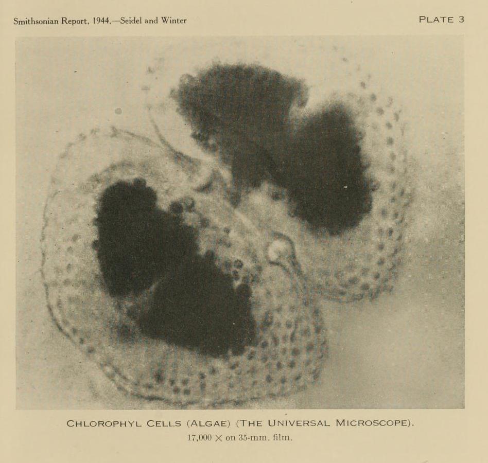
Possibly a diatom. Caption - "Chlorophyl cells (algae)" - doesn't make sense. From my limited knowledge of these things, it looks like a diatom, which seem to be a very commonplace subject for microscopes. But one article I read implied that a trained biologist had been very impressed by this photo - maybe the writer had the wrong end of the stick. Not sure what sort of diatom it is, if it is one.
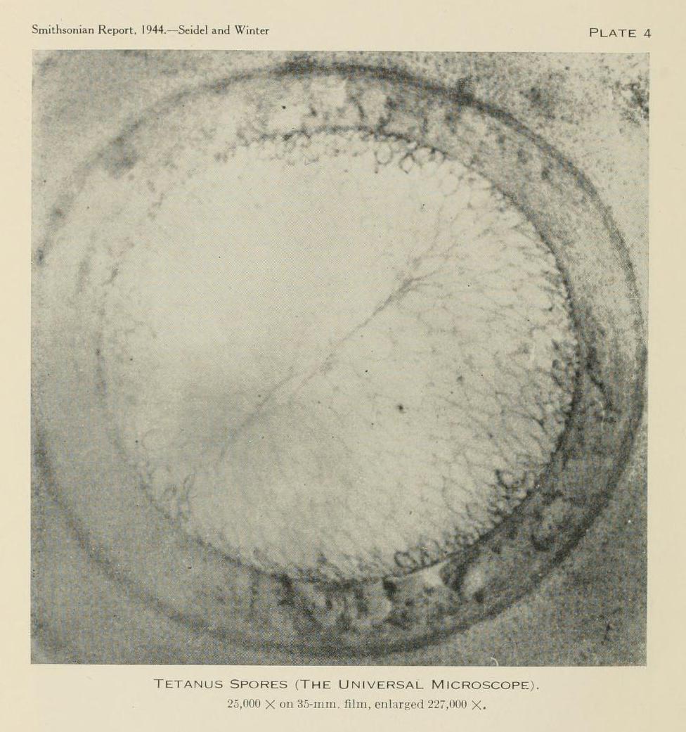
Tetanus spore Supposedly, the head of a tetanus bacterium, showing the spore, which, according to my brief Internet research, is possibly about 5 micrometres wide - so the quoted magnification of 23,000 sounds about true, if it really is a tetanus bacterium.

Typhoid bacterium Supposedly, a typhoid bacterium, which, according to my brief Internet research, is possibly about 1 micrometre wide by 3 long.
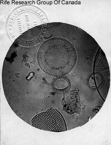
Dunno. Included because it's not labelled and I'm not sure what it is. Diatoms? No reference for where this one originally came from. This copy from https://www.rife.de/images/calibrate1.jpg.

$\endgroup$
3
  • $\begingroup$ "Magnification" isn't a particularly useful measure; you can magnify anything as far as you want as long as you don't care about resolution. $\endgroup$
    – Bryan Krause
    Commented Nov 16, 2022 at 15:13
  • $\begingroup$ Top picture is probably a dinoflagellate, possibly Gymnodinium family. Last are almost certainly diatoms of some sort, maybe Actinocyclus. Browse the beauty here $\endgroup$
    – bob1
    Commented Nov 16, 2022 at 23:43
  • $\begingroup$ @BryanKrause That's true - and the "typhoid bacterium" does seem to be magnified considerably further than it has the resolution for. Not sure how to express "resolution" numerically, though, so I just left it at "magnification", meaning "magnification possible without the image being reduced to a smudge". (Which, in the case of the "typhoid bacterium", might be about half what it has there - but making the image smaller might have made the caption illegible). $\endgroup$
    – A. B.
    Commented Nov 17, 2022 at 4:59

1 Answer 1

7
$\begingroup$

Having had a quick read of the report, I have a few comments - first off, this seems to be a second-hand report of something that hasn't actually been observed by the authors.

Second, the light microscopy resolution limit is limited by

$$wavelength/2$$

As defined by Abbe's law, where x,y is the plane of focus, $λ$ is wavelength and NA is the numerical aperture of the lens.

$$Abbe Resolution_x,_y = λ/2NA$$

Practically this means that the shorter the wavelength, the smaller the resolution can be. For visible light the shortest is around 380 nm, so the limit would be 190 nm or 0.19 micrometers. This is about the size of the smallest bacteria and a few viruses reach this size. However, because of the limitations in how we make optical media (lens glass etc) the practical limit is quite a bit higher. Electrons have very short wavelengths (around 4 picometres), so much greater magnifications can be achieved, which is why EM is used for imaging viruses. Based on these physical laws, I would be very surprised if Rife achieved the magnifications he thought he achieved. Indeed, many of his claims of discoveries based on his magnifications (e.g. that cancer is caused by tiny E. coli-like cells that take up to 5 morphologies) are utter nonsense - of course he was the only one capable of seeing these, with his high-power microscope and he wouldn't let anyone else handle or use these...

So, I do have doubts about the magnifications expressed. In the journal Rife's magnifications are based on "diameters" rather than an actual dimensionless magnification (ratio of actual size to apparent size). Diameters was a commonly used method of working out object sizes, but I don't know how to convert between the two. I would say, seeing as there are actual fundamental physical limits to the resolution, that what is being seen here are what is known as empty magnification - increase in size without increase in resolution. The same sort of effect you get with a digital camera when it doesn't have an optical zoom, but instead relies on making the pixels bigger in the field of view. In this case it will simply be bigger projection than the limits of resolution.

Now, on to the actual question. What are these things? Taking on face value that images 1-3 are what the captions say they are on pages 222-224 of OP's linked publication, then we have some candidates.

OPs first image is almost certainly a dinoflagellate, these are a polyphyletic group of single-celled algae that, like diatoms, have beautiful structured cases. They also have flagellae, which enable movement. Which dinoflagellate this is, I don't know, they are diverse and not at all my field. I suspect something in the Gymnodiniaceae. Dinoflagellates are about 30 micrometers in size, so easily within the limits of a light microscope, and you can see this in the ability to resolve the chloroplasts inside the organism. This image has no better resolution than would be produced by a decent conventional light microscope, only much bigger - camera trickery!

Image 2 could well be the spore end of Clostridium tetani, as the caption suggests. The spore is round and surrounded by a thick walled structure, but to my knowledge should have the rest of the bacterial body coming off to one side - looking rather like a spoon, with the body being the handle and the bowl of the spoon being the spore. I would expect to see the body of the bacterium sticking off to the side here, so I don't know for sure that it is what it is claimed to be, but we could be looking down on one along the longitudinal axis. There is some sponge-like "structure" visible in the middle of the "spore", but to me this looks like what you sometimes get in the middle of air bubbles on the slide.

Image 3 could be a member of the Salmonella group of organisms that cause typhoid, as these are slightly sausage shaped rod bacteria, however, I have seen many a stain blob that looks just like this using a low power objective. Note that Salmonella has flagellae too, and despite the large size and supposed massive magnification, we still can't see them. Hence, magnification and resolution probably not what is noted. So, whether this is actually Salmonella, is hard to say, but it could be.

Image 4 is from a different source - Rife.de, who seem to be an website for promoting Rife and his "cancer treatment". These look a lot like diatoms to me, but there is nothing to suggest that these were taken by Rife, and they look suspiciously like modern photographs using a modern microscope with something like Normarski optics or perhaps dark-field illumination, given that all the rest of Rife's images look much less clear.

$\endgroup$
3
  • $\begingroup$ Thanks very much for looking at this in so much detail! According to britannica.com/technology/magnification "diameters" is the same as "x", so, if so, that's easy at least! $\endgroup$
    – A. B.
    Commented Nov 17, 2022 at 6:37
  • $\begingroup$ I'm not sure where Rife.de get their images from, other than the caption "Rife Research Group Of Canada" (which isn't them). They seem to attempt to be a site seriously discussing the topic (there's a forum attached, and a lot of other archive materials), but they do seem to take the "it's basically real" position. Full set: rife.de/observations-with-the-rife-microscope.html . $\endgroup$
    – A. B.
    Commented Nov 17, 2022 at 6:45
  • $\begingroup$ In #3 the flagellae seem reasonably visible at the right, if not well focused. The internal detail is reasonably impressive - I'm not ready to hail it as a wonder, but it was at least a well-done job of staining and photography. $\endgroup$ Commented Nov 20, 2022 at 18:07

You must log in to answer this question.

Not the answer you're looking for? Browse other questions tagged .