5
$\begingroup$

There are variabilities in the heart rate as well as strength of heartbeats. I want to know if one can predict which heartbeat will be strong or weak based on the corresponding EKG/ECG signal. I am interested in the displacement of brain due to the heartbeat, which probably correlates with the peak pressure, and total volume caused by the beat.

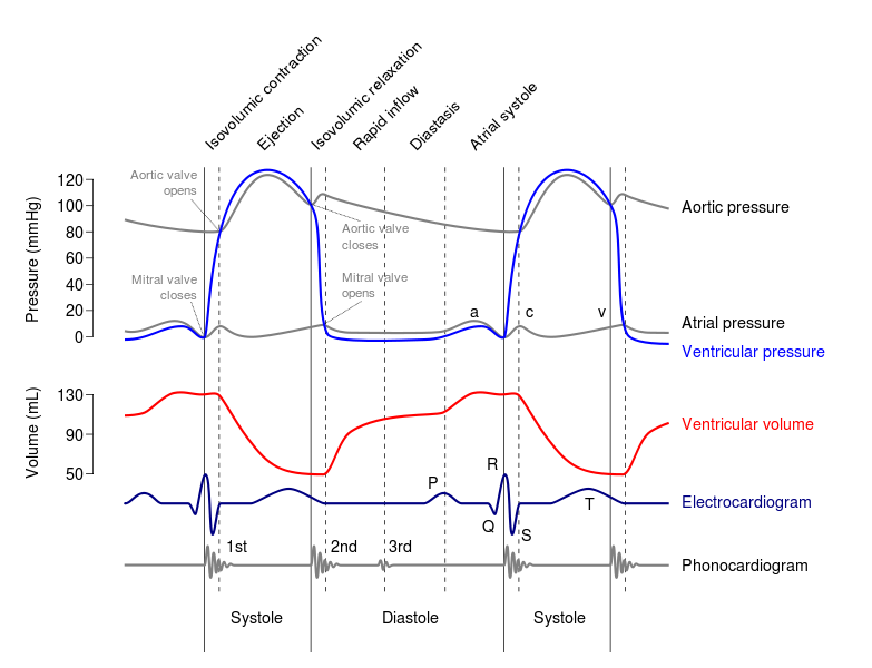
If there is such a correlation, which part of EKG would be related? The interval between P and R? The amplitude of QRS?

Human or primate information is preferred.

enter image description here image from wikipedia

$\endgroup$
5
  • $\begingroup$ Predict? I have no idea. I know V-tach and A-tach can be reasonably predicted by increasingly chaotic signals on an EKG, but I don't think the equipment would have the granularity to predict "strong" and "weak" beats. $\endgroup$
    – MCM
    Commented Aug 13, 2014 at 17:31
  • $\begingroup$ Are you referring to stroke volume, pulse pressure, or something else? $\endgroup$ Commented Aug 14, 2014 at 0:21
  • $\begingroup$ @louism Thanks for the interest. I suspect volume would be the best, but pressure or delay could also be useful. Any reference is welcome! $\endgroup$
    – Memming
    Commented Aug 14, 2014 at 17:23
  • $\begingroup$ @MCM to me it's not a matter of granularity, but fundamental plausibility. I can always improve signal-to-noise ratio, and do signal processing to improve the readings. But is there any signal related to beat strength at all? $\endgroup$
    – Memming
    Commented Aug 14, 2014 at 19:36
  • $\begingroup$ Is it plausible to estimate stroke volume from EKG (with significant measurement error)? Perhaps. Is it likely that you will achieve reasonable results without several months of research and access to large databases of patients who had both an EKG and a cardiac echo (gold standard for SV) in order to validate your results? Probably not. That said, if you did find a way, you would probably become incredibly rich. $\endgroup$ Commented Aug 16, 2014 at 20:20

3 Answers 3

3
$\begingroup$

Higher QRS complexes particularly in the lateral leads would suggest hypertrophy of the left ventricle. This in turn would suggest a stronger pressure is in place, although it is not known whether this is because it is required (incompetent valves) or a physiological response to exercise (where the volume is likely to actually be bigger).

Alternatively, if it really is just displacement of the brain you're interested in, why not use an EEG taken with an ECG and measure if there is a variation with each heartbeat. Then you can tease apart whether this is due to pressure or oxygenation or many other things.

Otherwise you could place something overlying the carotid which will loosely, but perhaps more accurately, reflect the pressure and one could potentially calculate the volume this way once enough other factors are known.

$\endgroup$
1
  • $\begingroup$ Thanks for the response. Hypertrophy would be a long term effect, right? I am interested in beat-to-beat variability. Do you have a reference for this by any chance? We are sure that it is the displacement of brain due to the heartbeat, and the displacement varies from beat-to-beat. We wish to control it as much as possible, but just wondering if there are better features in ECG that can be used to do so. $\endgroup$
    – Memming
    Commented Aug 15, 2014 at 13:53
3
$\begingroup$

It is unclear from your question what exactly "beat-to-beat variability" means from a physiological perspective. It would help if you could provide more information on the application you are working on and what kind of sensors you have access to.

If you wanted to measure the force of the arterial pulse, you would need to measure pulse pressure (which is the systolic blood pressure minus the diastolic blood pressure). However, these pressures do not only depend on the heart's action, but also on blood vessel compliance - which is influenced by ageaing, disease processes, and changes in the concentration of vasoactive substances in the blood. The EKG signal does not provide any of these latter parameters, and therefore it is highly unlikely that you would be able to estimate pulse pressure from an EKG. That being said, you could very easily measure pulse pressure with an arterial line, which would be the gold standard for such a task.

If you wanted to measure the force of the heartbeat, you could look at ejection fraction, which is a measure of how much blood is ejected from the ventricle in one heartbeat. Again, this is not related purely to the electrical activity of the heart (contractility and heart rate), but also to extra-cardiac parameters (preload and afterload). The EKG, which only looks at the intrinsic electrical activity of the heart, does not provide information on these extra-cardiac factors.

You could settle and look purely at cardiac contractility, which measures how much force the heart generates during a heartbeat. Keep in mind that this does not reflect how "well" the heart is working - you could have very good contractility, but a poor ejection fraction for other reasons. This is also not necessarily related to how "strongly" a person will feel his/her heart beating. I'm aware of one paper on how to measure cardiac contractility from EKG tracings.
$\endgroup$
3
  • $\begingroup$ Thank you for the detailed response! We have a setup where surface of cortex is imaged at high speed, and the image is contaminated by the displacement of the brain. This displacement significantly varies per beat, and we want to remove the effects. EKG and the video are pretty much all we are measuring. $\endgroup$
    – Memming
    Commented Aug 16, 2014 at 21:24
  • $\begingroup$ The paper you shared could be very useful. Thank you again. $\endgroup$
    – Memming
    Commented Aug 16, 2014 at 21:43
  • $\begingroup$ The displacement of the brain would be related to pulse pressure. I doubt you can measure this from an EKG signal. Again, the paper I shared shows a correlation between EKG signal and contractility, but moving from contractility to pulse pressure requires more information. $\endgroup$ Commented Aug 17, 2014 at 17:06
1
$\begingroup$

If you want a likely predictor candidate for the pulse wave amplitude (which seems to be what you are looking for), you will need to take into account some factors:

  • is the heart normal or pathologic?
  • are the vessels normal or pathologic?

Now from your question, I assume that you want your model to represent a sane vascular system. In this case, the best predictor of a strong heart beat would most likely be the area under the curve (AUC) of the QRS complex. However, you seem interested exclusively in the arterial pulse wave of the great circulation. If that is the case, Maybe it would be better to reduce your sampling to the QR waves AUC in an attempt to restrict your signal to the left ventricle contraction.

In short, you are trying to predict the beat-to-beat cardiac output from ECG. Some have already investigated that sort of thing. You could try looking at this device for a start, and then look up the list of supporting reference for additional information.

good luck!

$\endgroup$
2
  • $\begingroup$ Great suggestions. Yes, we are interested in the normal heart. I'll test with your suggestions. Thank you. $\endgroup$
    – Memming
    Commented Aug 18, 2014 at 17:19
  • $\begingroup$ @Memming BTW it seems that your question is just geared towards correction of other measurments. Can you give us a sample image or video of what your cortex camera is recording? Because it could be far better to try to adjust these images using fixed image features and compensating for the movements in code than trying to derive that from ECG... $\endgroup$
    – Raoul
    Commented Aug 18, 2014 at 20:03

You must log in to answer this question.

Not the answer you're looking for? Browse other questions tagged .