16
$\begingroup$

I have a technical question regarding the syntax in Protein Data Bank files. In the protein with PDB# 1AE9 (pdb file), there are two lines in the .pdb file:

HELIX    4   4 MET A 255  ILE A  265  1                                  11    

SHEET    2   B 2 ILE A 253  MET A 255 -1  N  ILE A 253   O  ILE A 248           

Now, as I understand the documentation, this means there is an alpha helix starting from residue "MET A 255" to the residue "ILE A 265". There is also a beta strand from "ILE A 253" to "MET A 255". We see that the alpha helix and beta strand share the residue "MET A 255".

The documentation describes these as:

  • "Name of the initial residue."
  • "Name of the terminal residue of the helix."

The wording suggests that the "initial" residue is included, but I'm unsure of whether or not the "terminal" residue would be included.

Question: Should I consider this residue as belonging to both the alpha helix and beta strand? Or should I consider the "terminal" residue as not belonging to the secondary structure element? (Or something else?)

$\endgroup$
1
  • 2
    $\begingroup$ ive seen records like this although it does not seem possible as alpha and beta have distinct ramachandran coordinates. i'll try to look at the structure and post an answer/opinion. a quick way around this is to find the phi/psi values for the amino acids in question and make the call yourself. $\endgroup$
    – shigeta
    Commented Aug 1, 2012 at 16:46

2 Answers 2

5
$\begingroup$

TLDR; Answer: You could consider this particular residue to belong to both structural elements, but it's a tricky call and depends on the method of secondary structure assignment.

Ambiguous secondary structure allocation comes up fairly often. Whilst obviously, not many people will be able to use this protein specifically, the below approach could be useful for other proteins.

Default pymol assignment

In PyMol I used fetch 1ae9 and highlighted M at position 255 on the A chain in red. I can see why this isn't a satisfying representation: the beta sheet allocation is very short, and the residue in question is clearly the beginning of a helix.
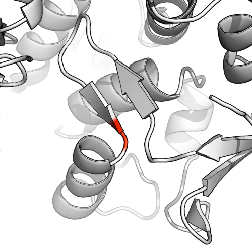
image showing that the residue is part of the beta sheet

DSS is more conservative

To run a more conservative secondary structure allocation run dss in pymol (watered down dssp). This reveals that the beta sheets were actually quite putative.
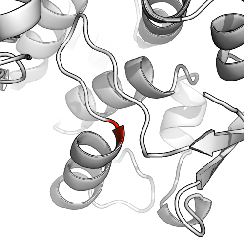
image showing that the residue is now part of the beginning of the helix

We can see why when we look at the stick model (below). There are only a few (2-4) residue pairs in proximity that would be available for hydrogen bonding, even assuming all 4 form H-bonds, calling this a bona fide beta sheet could be seen as a bit generous.
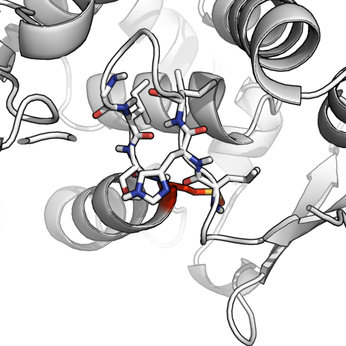
image showing the stick model.

Dual assignment can be okay.

Looking at this example (1ae9), we see the potential for H-bonding with another putative beta sheet, as well as a backbone angle that starts to form a helix. It's a classic case of ambiguous dual assignment. @AlexanderDScouras draws a reasonable conclusion that both is okay and to directly answer the question: dual assignment is possible, and allowed. I would prefer to rule out beta sheets in this particular case, but on the condition that one highlights that there is plenty of hydrogen bonding in the hairpin loop.

So long as it's reasonable.

So long as you can reasonably justify the assignment it's probably okay. You can manually assign any residue and secondary structure.

# set residue 155 to be alpha-helical
alter 155/, ss='H'

# update the scene in PyMOL to reflect the changes.
rebuild

Evidence is always a good idea to support manual assignments. A quick and thorough way would be to run the sequence through a few secondary structure predictors, or use the genesilico metaserver to save yourself some hassle (you'll need an account) (this does feel like going back a step since you now have a structure, so exercise caution - if the sequence prediction doesn't look correct according to the structure, it's probably not). Another method as suggested by shigeta is running this through a Ramachandran plot (RAMPAGE is a favourite).
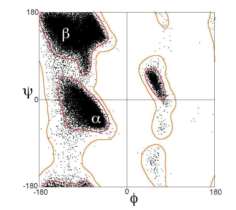
Ramachandran plot showing discreet cut-offs between alpha and beta

If it's very very unclear then discuss it thoroughly and clearly whenever this part of the protein is relevant. These secondary structure assignments have somewhat arbitrary cut-offs after-all, and when things get near the thresholds, it's important to approach the situation with clarity and specificity.


Ramachandran plot image source: By Dcrjsr - Own work, CC BY 3.0, https://commons.wikimedia.org/w/index.php?curid=9105708

$\endgroup$
2
  • $\begingroup$ A most informative answer! But I'm still surprised PDB allows such overlapping secondary structure markup. Perhaps their attitude is that all that is really important are the coordinates — it's up to the individual how they choose to visualize it. $\endgroup$
    – David
    Commented May 28, 2016 at 9:33
  • 1
    $\begingroup$ @David Precisely. The terms α and β are there to help us quickly talk about broad biochemically similar parts of proteins. When the biochemistry get's stretched to the limits of the definition, the biochemical similarities become more vague and perhaps it's time to talk about the specific biochemistry in that protein again. There will be times when this residue example acts more like a helix, and other time when it is more like a sheet. $\endgroup$
    – James
    Commented Jun 1, 2016 at 1:42
4
$\begingroup$

This is almost more a philosophical question about how you would like to define helices and sheets, which I would argue is not so well defined. shigeta mentions that they have well defined ramachandran coordinates, but that's for the central residues. Terminal residues are far more flexible. The more traditional definition is along the lines of the DSSP algorithm, i.e. what is their main-chain hydrogen bonding pattern.

The DSSP algorithm is a bit conservative about assignments, often leaving out the first and last residues in both sheets and helices. In deposited structures, you can compare author annotations to DSSP and STRIDE and see the authors will often assign longer runs than either. And I have certainly seen structures where a residue is annotated as both, especially when it lies at the junction between two secondary structure elements.

Looking at the structure, residues 254 and 255 are clearly hydrogen bonding into a beta-sheet on one side and an alpha helix on the other, so I see no reason they shouldn't be annotated into both.

$\endgroup$

You must log in to answer this question.

Not the answer you're looking for? Browse other questions tagged .