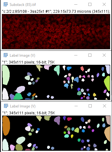
I've got some images such as the one in this post, obtained from the neuroepithelium of a chick embryo in a confocal microscope, where nuclei have been fluorescently labeled. I'd like to be able to segment these nuclei, in particular to see their total area (which means I don't necessarily need them to be separated) and to count them. I tried using StarDist on Fiji, but I've obtained some very bad results, with it detecting basically no nuclei.
Does anyone have any suggestions on what I might do?
Here's a link to the original image in tif format: https://drive.google.com/file/d/1GqsUI-xnTPSKnRv_ECxJL7K785_qbq2c/view?usp=sharing
Below there's a comparison of my original image(above), with the StarDist output with low probability/score threshold and low overlap threshold (middle) and with default settings (below). Previously, default settings would output a completely black image, but it seems to have changed.


