2
$\begingroup$

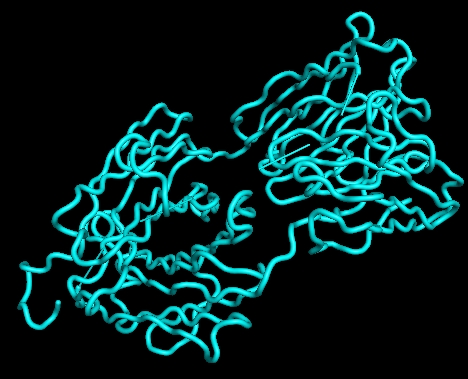
I am using pymol to visualise the secondary structure of protein using its cartoon representation. The pdb file comes from a simulation, which contains multiple frames. After loading the pdb, its secondary structure (e.g. sheet, helix) could not been recognised. Surprisingly, if only one frame is kept, its secondary structure could be seen. So how to enable secondary structure recognition for a pdb file with multiple frames?

An example of the pdb file with multiple frames is shown below.

REMARK    GENERATED BY TRJCONV
TITLE     Protein in water t=   0.00000
REMARK    THIS IS A SIMULATION BOX
CRYST1  116.453  116.453  116.453  90.00  90.00  90.00 P 1           1
MODEL        1
ATOM      1  N   ASP     1      85.582  59.777  48.367  1.000.0000           N
ATOM      2  H1  ASP     1      84.882  59.067  48.507  1.000.0000           H
ATOM      3  H2  ASP     1      85.162  60.617  48.747  1.000.0000           H
... ...
ATOM   6615  OT  ALA   442      28.032  36.877  69.157  1.000.0000           O
ATOM   6616  O   ALA   442      30.092  36.087  68.677  1.000.0000           O
ATOM   6617  HO  ALA   442      30.072  35.867  69.597  1.000.0000           H
TER
ENDMDL
REMARK    GENERATED BY TRJCONV
TITLE     Protein in water t= 1000.00000
REMARK    THIS IS A SIMULATION BOX
CRYST1  116.384  116.384  116.384  90.00  90.00  90.00 P 1           1
MODEL        2
ATOM      1  N   ASP     1      75.052  41.097  56.132  1.000.0000           N
ATOM      2  H1  ASP     1      75.622  41.407  55.352  1.000.0000           H
ATOM      3  H2  ASP     1      75.602  41.357  56.932  1.000.0000           H
ATOM      4  H3  ASP     1      74.682  40.167  56.272  1.000.0000           H
ATOM      5  CA  ASP     1      74.032  42.157  56.202  1.000.0000           C
... ...
ATOM   6615  OT  ALA   442      45.292  49.247  90.922  1.000.0000           O
ATOM   6616  O   ALA   442      47.102  49.617  89.632  1.000.0000           O
ATOM   6617  HO  ALA   442      47.662  49.327  90.342  1.000.0000           H
TER
ENDMDL

enter image description here enter image description here

$\endgroup$
8
  • $\begingroup$ I suspect that the PDB is malformed. Is it available online somewhere so I can take a look at it? Alternatively, could you post and excerpt of the HELIX and/or SHEET records from the file? $\endgroup$
    – canadianer
    Commented Feb 2, 2018 at 18:52
  • $\begingroup$ I notice that the ATOM records don't contain a chain identifier. I'm not sure if that would prevent PyMol from recognizing the secondary structure elements. $\endgroup$
    – canadianer
    Commented Feb 2, 2018 at 18:59
  • 1
    $\begingroup$ @canadianer the chain ID is not the problem, as it works for a single frame $\endgroup$
    – lanselibai
    Commented Feb 2, 2018 at 19:06
  • $\begingroup$ Okay... Does an NMR model with multiple conformers display correctly (5VKV, for example)? $\endgroup$
    – canadianer
    Commented Feb 2, 2018 at 19:10
  • $\begingroup$ So it must be this particular PDB. If there's anything wrong with the portion of the file you've posted, I don't know what it is. Does the file with multiple models have HELIX/SHEET records? $\endgroup$
    – canadianer
    Commented Feb 2, 2018 at 19:20

1 Answer 1

2
$\begingroup$

It seems that if no HELIX record is present in the PDB, PyMol attempts to assign secondary structure itself. For whatever reason, it does not do this if multiple MODELs are present. You should be able to use the dss command to force PyMol to calculate secondary structure in a PDB with multiple MODELs.

$\endgroup$
2
  • $\begingroup$ Thanks for this, it was so easy to get this "fixed"! Works like charm. $\endgroup$ Commented Nov 18, 2021 at 9:44
  • $\begingroup$ @MarcinMagnus You’re welcome! $\endgroup$
    – canadianer
    Commented Nov 18, 2021 at 16:56

You must log in to answer this question.

Not the answer you're looking for? Browse other questions tagged .