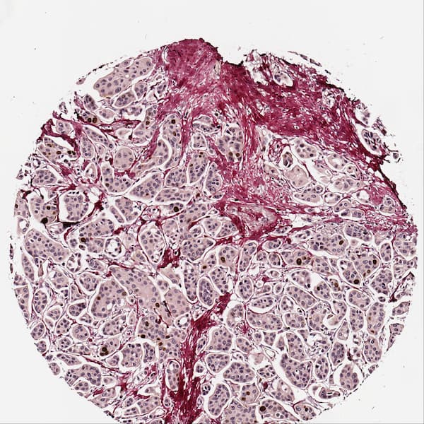
I am working on a project which involves writing computer software to analyse histological images. A typical image looks like this:
It is a Hematoxylin and Eosin stained biopsy of breast cancer tissue. Being a programmer without biology background, I would like to get insight into how a pathologist analyses such images.
More specifically:
- Which cells are cancer cells and which are regular cells?
- Can an invasive margin (a curve marking the boundary of cancer invasion into the tissue) be seen in this image?
- Can a pathologist provide a TNM classification looking at the image? If yes, what would be the T, N, M, G values for this image?

