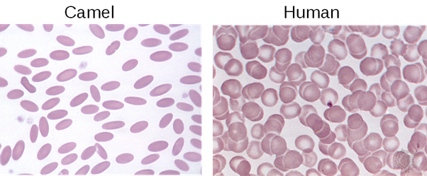
Unlike humans and most of mammals, which have a biconcave (or "doughnut shaped") RBC, camels have oval shaped (or elliptical) erythrocytes.
The very first result in Google shows this:

That feature (which protects them from dehydration) is not unique to camels, but occurs in other Camelidae as well. This is an image of Llama blood:

Those are reliable sources of images (optical microscopy) of real cells.
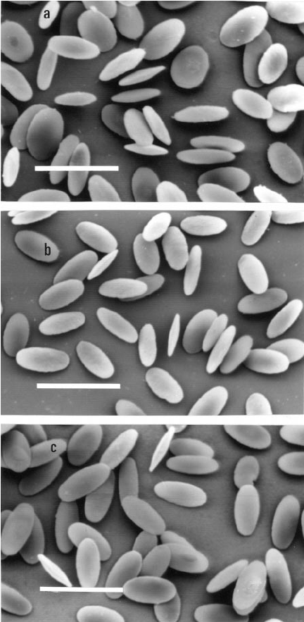
However, for your project, I believe a scanning electron micrograph can better show the shape.
This is an SEM of elliptical camel (a) and alpaca, another Camelidae (b and c), erythrocytes:

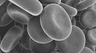
For comparison, this is the biconcave human erythrocyte:

PS: There is this misinformation that camels' erythrocytes are nucleate. You can read this in several web pages. I have no idea who made this up, or why people keep sharing this wrong information: camels, like any mammal, have anucleate erythrocytes. See the post Do camels have nucleated RBCs or enucleated RBCs? for more information.
EDIT: The way your question is worded it really seems that you're just asking about the reliability of those sources. However, according to your comment below, it seems that you are indeed looking for better images. It's hard finding better ones than those indexed by Google, but since your goal is modelling those cells, here are two papers (about a protozoan parasite of camelids) which has some SEM that may suit you. The images are not high-resolution, but the cells are shown in several angles:
In both papers the SEMs are at the bottom (just skip the optical ones, they are not good).


