31
$\begingroup$

I am not quite sure if it is true, but I read somewhere that within 7 years all the body's cells are replaced with new ones. I am not quite sure if it is cells or atoms.

If it is then why do tattoos persist for so long? If the cells which were impregnated with the dye have been replaced, then why do the tattoos still remain?

$\endgroup$
3
  • 2
    $\begingroup$ That's not true - some cells turn over quite quickly, others take months or years, others never replicate. $\endgroup$
    – MattDMo
    Commented Aug 9, 2015 at 6:20
  • $\begingroup$ It's a damn pity that that's not how it works, wouldn't have minded getting a tattoo if I knew it would fade in 5 years time and have disappeared entirely in another 10. $\endgroup$ Commented Aug 9, 2015 at 16:07
  • 1
    $\begingroup$ Smarter Every Day explained this quite well on their YouTube channel. $\endgroup$
    – Ajedi32
    Commented Aug 10, 2015 at 14:57

2 Answers 2

29
$\begingroup$

First, tattoo pigment isn't injected into cells. If you were to puncture a cell with something the size of a tattoo needle, it would die - full stop. Many cells are destroyed in the process though, mostly by tearing, which initiates the wound healing process.

So what actually happens is the particles lodge in between the cells of the dermis (the layer below the epidermis, or outer layer of skin) and new cells crowd around it as damaged cells are replaced.

Since the particles in good tattoo ink are too big to be carried off by macrophages, they just sit there. That doesn't stop them from trying, though. Macrophages take up the pigment but are unable to escape with their garbage. Fibroblasts envelope the particles, both intracellularly and within the extracellular matrix they generate. At the end of a fibroblast's life, its contents are taken up by the same process - still too big to remove.

Smaller particles will be carried off by macrophages capable of both consuming the particles and migrating into the lyphatic system, which is why some ink fades more than other kinds. Ink composed of fine particles would be expected to fade severely. New tattoos are also sharper because some of the ink that was injected is lodged in the epidermis, which is the layer that grows out and is shed over time.

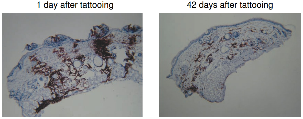
In this biopsy of a tattooed mouse, you can see how the epidermis has carried some of the pigment out of the skin while much of the dermal layer's pigment has been either removed lymphatically or enveloped:

Mouse tattoo biopsy

Source: Tattooing of skin results in transportation and light-induced decomposition of tattoo pigments--a first quantification in vivo using a mouse model.

Thus, over time the pigment will be moved around a bit by this cellular activity, in fact deeper into the dermal layer on the whole.

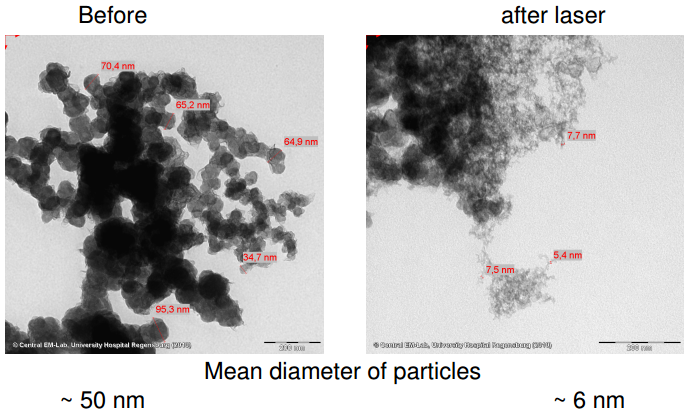
The intent of using laser light to remove tattoos is to break those particles down into smaller pieces burst any cells containing them - again initiating the wound healing process. However, unlike in previous cases, when macrophages reach the site they can now sweep the remaining particles away to the lymph nodes.

Pigment particles before and after lasing

Sources

Secondary sources:

$\endgroup$
6
  • 8
    $\begingroup$ It would be great if you could add citations because this does not sound quite intuitive. For instance, when you mention particle size and clearance from dermis has a connection, then add a reference for that. Also, you should add some reference for the particle size distribution of the tattoo ink. Most tattoo pigments are composed of metal salts, there is also a possibility of leaching (I am not sure how it does not happen. There are some reports on diffusion of tattoo pigment though) $\endgroup$
    – WYSIWYG
    Commented Aug 9, 2015 at 14:11
  • $\begingroup$ Additionally, it is very strange to phrase the sentence "puncture a cell with a tattoo needle" when the tattoo needle's tip is hundreds of times larger than a cell. $\endgroup$
    – March Ho
    Commented Aug 9, 2015 at 15:23
  • 1
    $\begingroup$ Destin produced a quite nice video that could be used as a reference for this answer. youtube.com/watch?v=D0B7F5UbTOQ $\endgroup$
    – wigy
    Commented Aug 9, 2015 at 19:06
  • 2
    $\begingroup$ -1 See my comment to March Ho's answer: the majority of the ink seems to be intracellular, albeit after a certain time. In any case, all the articles that I've read say there is no ink in the epidermis. $\endgroup$
    – biozic
    Commented Aug 9, 2015 at 20:48
  • 1
    $\begingroup$ @MarchHo Some people aren't familiar with the relative sizes of cells and needles, so I thought that would be important to mention. $\endgroup$
    – jzx
    Commented Aug 9, 2015 at 23:54
12
$\begingroup$

Contrary to the other answer posted, this paper shows via microscope images that the tattoo ink is in fact absorbed into cells, and forms small intracellular round granules.

Electron microscopy of untreated tattoos revealed membrane-bound pigment granules, predominantly within fibroblasts and macrophages, and occasionally in mast cells. These granules contained pigment particles.
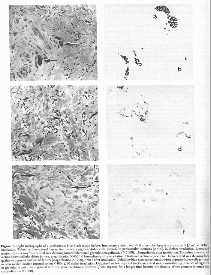
enter image description here Images a, c and e are images of toluidine blue stained cells, while images b, d and f are images of unstained cells showing the tattoo pigments.

Images a and b are prior to the laser treatment, c and d directly after the treatment, and e and f 90 days post treatment with the laser.

The reason why the tattoo marks persist is not because the pigments are deposited extracellularly, but that they are deposited intracellularly.

The pigments form intracellular granules that are not broken down, and therefore in the absence of external forces such as a laser, the pigments will remain there for long periods of time.

This paper (thanks to biozic) also describes the tattoo ink being persistently found intracellularly instead of extracellularly.

Biopsies obtained from tattoos 1, 2, 3, and 40 years old differed only in the types of ink used. All the ink particles were found to be located in dermal cells. The epidermis was completely devoid of pigment particles. The basement membrane was continuous at the epidermal-dermal junction. Ink particles were found throughout the upper dermis but all were within the boundary of a cell membrane.

According to this TED video (which unfortunately does not state its primary sources), the fibroblasts that contain these engulfed ink particles are themselves taken up by newer fibroblasts when they die, therefore the ink particles remain in the dermis and are not removed by cellular renewal.

$\endgroup$
5
  • 2
    $\begingroup$ Several other articles confirm that the majority of ink particle are intracellular (mainly in fibroblasts and macrophage) after the skin healing process is done [dx.doi.org/10.1111/j.1365-4362.1987.tb00590.x]. But, according to these articles, pigments are not deposited into cells: they end up there after a complex and time-consuming process. $\endgroup$
    – biozic
    Commented Aug 9, 2015 at 20:33
  • $\begingroup$ @biozic Can you point out where in the article does it say that pigments are not deposited into cells? The 24 hour post tattoo result clearly describes intracellular tattoo ink deposits. $\endgroup$
    – March Ho
    Commented Aug 9, 2015 at 22:54
  • $\begingroup$ Do these cells persist for a long time? Mast cells release their granules when stimulated and macrophages are chemotactic (can move around). Only fibroblasts seem static but even they can undergo apoptosis. So as the OP asked, what would happen when these cells die/move and are replaced by new cells? $\endgroup$
    – WYSIWYG
    Commented Aug 10, 2015 at 4:32
  • $\begingroup$ @WYSIWYG See the video citation. I was unable to find a better primary source. $\endgroup$
    – March Ho
    Commented Aug 10, 2015 at 4:40
  • $\begingroup$ @MarchHo What I wanted to say is that ink particles aren't specifically deposited into cells. According to the article, when they looked at a 24h-aged tattoo, "Homogenization of the epidermis, the epidermal-dermal junction, and the upper portion of the dermis was almost complete". "Both epidermal and inflammatory cells had ink particles in their respective cytoplasms [...] Ink particles were also found in the enlarged intercellular spaces". $\endgroup$
    – biozic
    Commented Aug 10, 2015 at 7:43

You must log in to answer this question.

Not the answer you're looking for? Browse other questions tagged .