what does the "two photon" means?
Ordinary confocal microscopy uses single photon of laser light to excite the molecule of fluorescent dye.
In two photon microscopy you use two photons, with lower energy, which cannot excite the molecule of dye on their own, but their interference in a specific place leads to summation of their energies and to excitation.
This has benefits such as decreased phototoxicity and increased precision (resolution) especially in z-plane.
how does the implement look like?
Does all relevant (genetically engineered) neurons light in the same color?
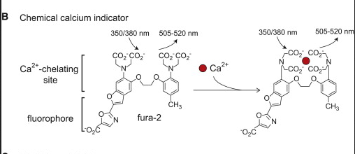
There are two types of fluorescent dyes for 2-photon Ca2+ imagining. Since you want to measure local concentration of Ca2+, you can either use a chemical which chelates the calcium ions and changes its conformation and fluorescent properties based on that e.g. fura-2.
Alternatively, you can prepare fused protein from calmodulin ( CALcium MODULated proteIN) and some fluorescent protein e.g. GFP and transfect the cells with this product.
This means you can transfect different cells with different GFPs and get different colors. Depends on the actual experiment design.
 Source
Is it the color that is measured or the intensity of the color?
Is it sensitive to absolute voltage or voltage changes?
You can analyse fluorescence intensity changes. The derived fluorescence intensities and ratios are plotted against calibrated values for known Ca2+ levels to learn actual Ca2+ concentration. Source
Is the purpose of this technique single neurons or brain paths?
Actually, with a good interpretation of the data it can be used for analysis of both.
For more information about calcium imaging, see for example: http://www.cell.com/neuron/fulltext/S0896-6273(12)00172-9
or http://www.pnas.org/content/100/12/7319.full
or corresponding wiki articles:
https://en.wikipedia.org/wiki/Two-photon_excitation_microscopy
https://en.wikipedia.org/wiki/Calcium_imaging

